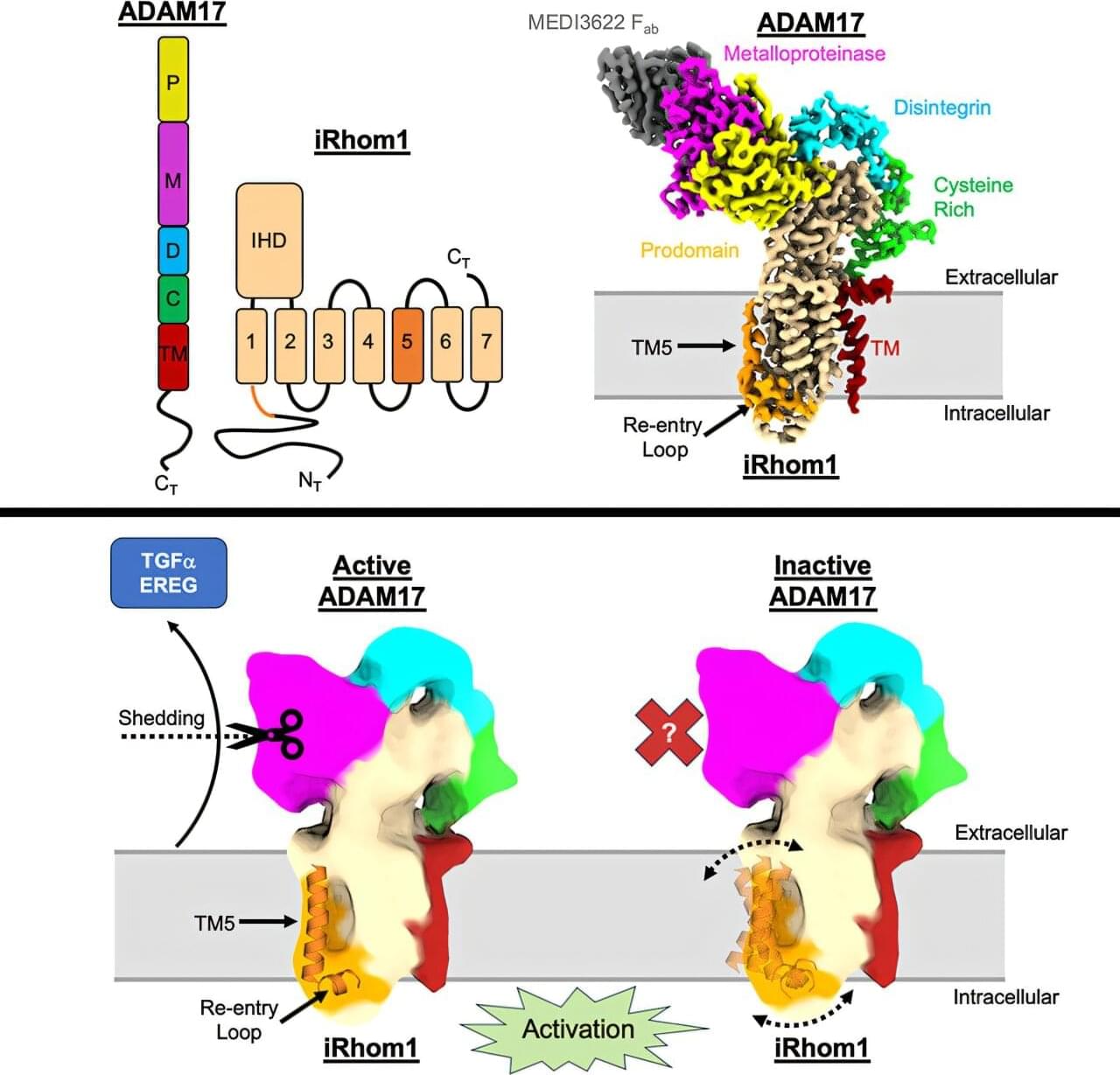
Toggle light / dark theme

OSAKA — A Japanese research team is making progress on the development of a groundbreaking medication that may allow people to grow new teeth, with clinical trials set to begin in July 2024.

The tooth regrowth medicine is intended for people who lack a full set of adult teeth due to congenital factors. The team is aiming to have it ready for general use in 2030.

In prior animal experiments, the medicine prompted the growth of “third-generation” teeth following baby teeth and then permanent adult teeth.

S dream. I.

Photodynamic therapy uses a drug that is activated by light, called a photosensitizer or photosensitizing agent, to kill cancer cells. The light can come from a laser or other source, such as LEDs. Photodynamic therapy is also called PDT.

Photodynamic therapy is most often used as a local treatment, which means it treats a specific part of the body.

The study, “Endothelial TDP-43 Depletion Disrupts Core Blood-Brain Barrier Pathways in Neurodegeneration,” was published on March 14, 2025. The lead author, Omar Moustafa Fathy, an MD/Ph. D. candidate at the Center for Vascular Biology at UConn School of Medicine, conducted the research in the laboratory of senior author Dr. Patrick A. Murphy, associate professor and newly appointed interim director of the Center for Vascular Biology. The study was carried out in collaboration with Dr. Riqiang Yan, a leading expert in Alzheimer’s disease and neurodegeneration research.

This work provides a novel and significant exploration of how vascular dysfunction contributes to neurodegenerative diseases, exemplifying the powerful collaboration between the Center for Vascular Biology and the Department of Neuroscience. While clinical evidence has long suggested that blood-brain barrier (BBB) dysfunction plays a role in neurodegeneration, the specific contribution of endothelial cells remained unclear. The BBB serves as a critical protective barrier, shielding the brain from circulating factors that could cause inflammation and dysfunction. Though multiple cell types contribute to its function, endothelial cells—the inner lining of blood vessels—are its principal component.

“It is often said in the field that ‘we are only as old as our arteries’. Across diseases we are learning the importance of the endothelium. I had no doubt the same would be true in neurodegeneration, but seeing what these cells were doing was a critical first step,” says Murphy.

Omar, Murphy, and their team tackled a key challenge: endothelial cells are rare and difficult to isolate from tissues, making it even harder to analyze the molecular pathways involved in neurodegeneration.

To overcome this, they developed an innovative approach to enrich these cells from frozen tissues stored in a large NIH-sponsored biobank. They then applied inCITE-seq, a cutting-edge method that enables direct measurement of protein-level signaling responses in single cells—marking its first-ever use in human tissues.

This breakthrough led to a striking discovery: endothelial cells from three different neurodegenerative diseases—Alzheimer’s disease (AD), amyotrophic lateral sclerosis (ALS), and frontotemporal dementia (FTD)—shared fundamental similarities that set them apart from the endothelium in healthy aging. A key finding was the depletion of TDP-43, an RNA-binding protein genetically linked to ALS-FTD and commonly disrupted in AD. Until now, research has focused primarily on neurons, but this study highlights a previously unrecognized dysfunction in endothelial cells.

“It’s easy to think of blood vessels as passive pipelines, but our findings challenge that view,” says Omar. “Across multiple neurodegenerative diseases, we see strikingly similar vascular changes, suggesting that the vasculature isn’t just collateral damage—it’s actively shaping disease progression. Recognizing these commonalities opens the door to new therapeutic possibilities that target the vasculature itself.”

Northwestern Medicine investigators have identified novel mechanisms regulating the development of the spinal column during embryonic development, findings that could inform new treatments for congenital scoliosis and other related birth defects, according to a recent study published in Nature Communications.

The spinal column of all vertebrate species, including humans, is divided into segments (vertebral discs), which give the spine both flexibility and mobility.

During early , these discs develop from specialized cells called somites and are sequentially “sliced” into separate discs, a process driven by a called the vertebrate segmentation clock.

For the first time, scientists have acquired direct evidence of rare, pulsing pear-shaped structures within atomic nuclei of the rare-earth element gadolinium, thanks to new research led by the University of Surrey, the National Physical Laboratory (NPL) and the IFIN-HH research institute in Bucharest, Romania.

The study, published in Physical Review Letters, provides definitive proof of a strong collective “octupole excitation” in the nucleus of gadolinium-150, a long-lived radioactive isotope of this rare-earth element, which is used in applications such as superconductors, nuclear power operations and MRI contrast materials.

The experimental signature is interpreted as the protons and neutrons inside the atomic nucleus vibrating in a coordinated pattern, resulting in a pulsing, asymmetric, pear-shaped structure.

One of the most enduring questions humans have is how long we’re going to live. With this comes the question of how much of our lifespan is shaped by our environment and choices, and how much is predetermined by our genes.

A study recently published in the prestigious journal Nature Medicine has attempted for the first time to quantify the relative contributions of our environment and lifestyle versus our genetics in how we age and how long we live.

The findings were striking, suggesting our environment and lifestyle play a much greater role than our genes in determining our longevity.

An interesting article where Lee et al. develop a new chemical label for studying the dynamics of select glycolipids found in tuberculosis bacteria. They target specific types of glycolipids that are involved in pathogenesis, opening the door to new insights on tuberculosis. As tuberculosis kills more than a million people every year, tools for studying the disease are sorely needed. #chemicalbiology #chemistry #microbiology

Central sensitization: analysis by physio meets science.

Neurophysiological Mechanism of Central Sensitization in the Spinal Cord following Surgery:

▶️ Central sensitization was first described by Woolf in 1983 (https://pubmed.ncbi.nlm.nih.gov/6656869/) as a form of long-term adaptive neuroplasticity that amplifies the transmission of nociceptive information by affecting spinal cord neurons and is believed to be a principal neurophysiological mechanism with regard to pain persistence.

▶️ Peripheral nociception can trigger a prolonged increase in the excitability of dorsal root ganglia (DRG) neurons, which transmit nociceptive signals to the spinal cord, resulting in central sensitization.

▶️ This condition involves heightened responsiveness of spinal neurons, driven by signaling molecules like adenosine triphosphate (ATP) and neurotransmitters such as glutamate (Glu) and substance P (SP).

▶️ These molecules activate specific receptors on spinal neurons, including purinergic receptor 2 (P2-R), N-methyl-D-aspartate receptor (NMDAR), α-amino-3-hydroxy-5-methyl-4-isoxazolepropionic acid receptor (AMPAR), and neurokinin 1 receptor (NK1R).

▶️ The activation of these receptors sets off a cascade of intracellular pathways involving enzymes like calcium/calmodulin-dependent protein kinase II (CaMKII), protein kinase C (PKC), protein kinase A (PKA), mechanistic target of rapamycin (mTOR), phosphoinositide 3-kinase (PI3K), and extracellular signal-regulated kinases 1/2 (ERK1/2), all of which amplify the transmission of nociceptive signals to the brain.