Toggle light / dark theme

PET scans reveal early signs of Parkinson’s and Lewy body disorders

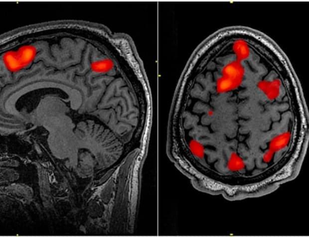
In a comprehensive Genomic Press perspective article published today, researchers from Fudan University and Shanghai University of Traditional Chinese Medicine have highlighted remarkable advances in the development of positron emission tomography (PET) tracers capable of visualizing α-synuclein aggregates in the brains of patients with Parkinson’s disease and related disorders.

The abnormal accumulation of α-synuclein protein is a defining pathological feature of several neurodegenerative conditions collectively known as synucleinopathies, including Parkinson’s disease (PD), multiple system atrophy (MSA), and dementia with Lewy bodies (DLB). Until recently, confirming the presence of these protein aggregates required post-mortem examination, severely limiting early diagnosis and treatment monitoring capabilities.

“The ability to visualize these protein aggregates in living patients represents a significant leap forward in neurodegenerative disease research,” explains Dr. Fang Xie, corresponding author and researcher at the Department of Nuclear Medicine & PET Center at Huashan Hospital, Fudan University.
Skin-based test could improve diagnosis of debilitating neurodegenerative disease

Researchers at the University Health Network (UHN) and the University of Toronto have developed a skin-based test that can detect signature features of progressive supranuclear palsy (PSP), a rare neurodegenerative disease that affects body movements, including walking, balance and swallowing.

The test, which the researchers describe in a recent issue of JAMA Neurology, could allow for more accurate and faster PSP diagnosis than current methods.

“This is important for assigning patients to the correct , but it will be even more important in the future as researchers develop targeted, precision treatments for PSP,” says Ivan Martinez-Valbuena, a scientific associate at the Rossy Progressive Supranuclear Palsy Centre at the UHN’s Krembil Brain Institute and U of T’s Tanz Centre for Research in Neurodegenerative Diseases.

Clinical features of Rathke’s cleft cyst with secondary hypophysitis and outcomes of endoscopic transnasal surgery versus features of common Rathke’s cleft cysts: a single-center retrospective cohort study

The aim of this study was to examine the clinical characteristics of Rathke’s cleft cyst (RCC) with secondary hypophysitis and compare them with the clinical characteristics of common RCC.

This single-center retrospective cohort study included cases of pituitary disease in which endoscopic transnasal surgery was performed from January 2011 to March 2023. Patients with RCC were identified, and secondary hypophysitis was subsequently identified based on pathological and MRI findings. Pathologically, the presence of lymphocytic infiltration into the normal anterior pituitary gland was used as a criterion for determining hypophysitis. On MRI, RCCs showing marked thickening of the cyst wall and pituitary stalk swelling ≥ 3.5 mm were considered as hypophysitis. A comparative study was performed at our institution using retrospectively collected data on RCCs with secondary hypophysitis and common RCCs.

The study included 11 patients with RCC with secondary hypophysitis (median age 36 years) and 95 patients with common RCC (median age 51 years). The proportions of patients with headache (90.9% vs 48.4%, p = 0.009), fever (63.6% vs 1.1%, p < 0.001), panhypopituitarism (90.9% vs 24.2%, p < 0.001), and diabetes insipidus (90.9% vs 21.1%, p < 0.001) were significantly higher in the RCC with secondary hypophysitis group than the common RCC group. Although reaccumulation rates and the time to reaccumulation did not differ between the groups, the operative rate when reaccumulation occurred was significantly higher in patients with RCC with secondary hypophysitis than in those with common RCC (75% vs 13%, p = 0.015).

Somatosensory Mapping Using a Novel Sensory Discrimination… : Operative Neurosurgery

Responses, necessitating refined somatosensory mapping techniques.

METHODS:

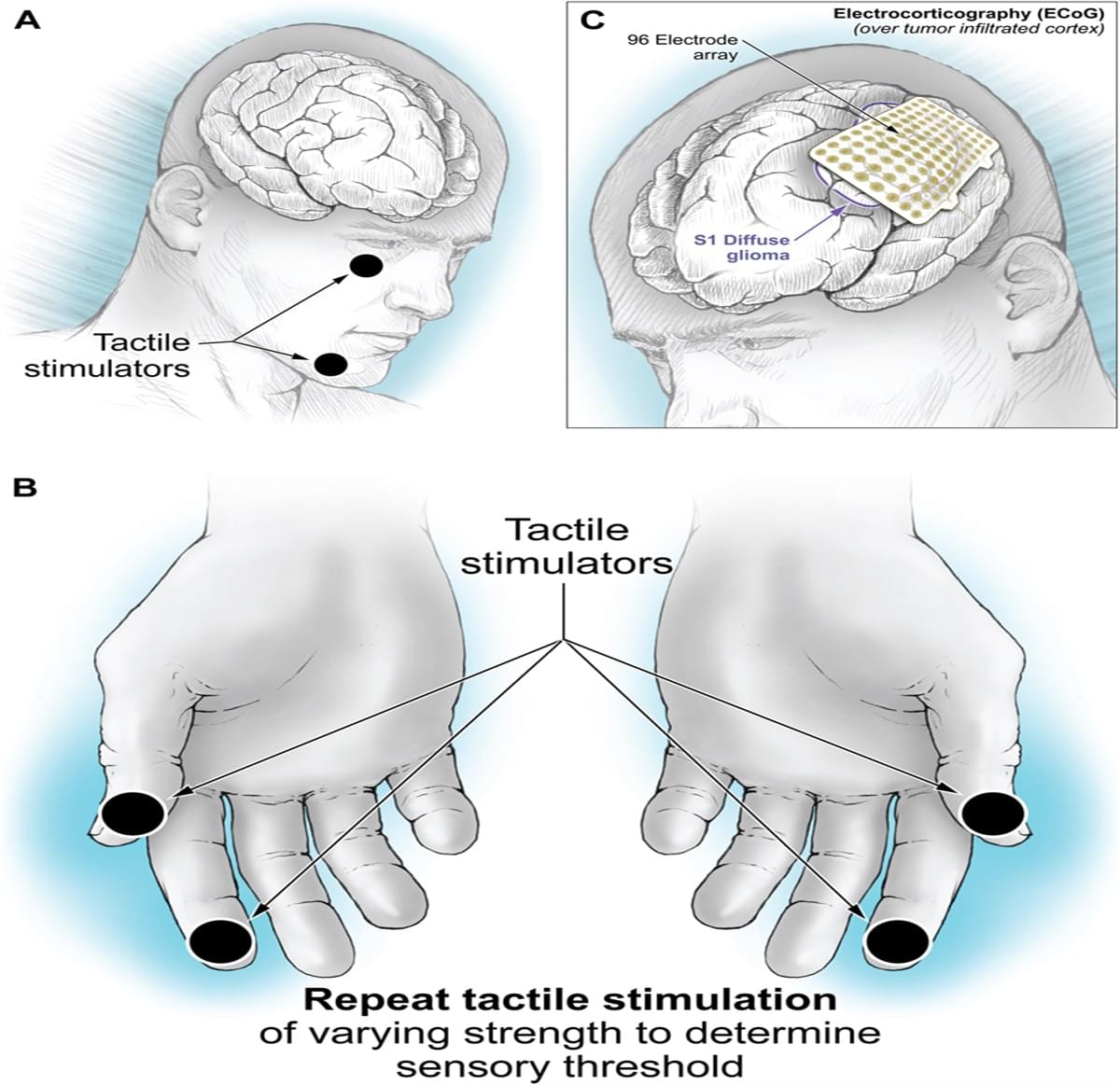
Using piezoelectric tactile stimulators on patients’ faces and hands, we delivered 25 Hz vibrations and prompted patients to discriminate between dermatomes. Testing included areas contralateral to tumor-infiltrated and to non–tumor-infiltrated cortical regions. Sensory thresholds were determined by reducing stimulus intensity based on performance. Intraoperatively, electrocorticography electrode arrays were used to map sensory responses, and postoperative assessments evaluated sensory outcomes.

New technique protects ‘architecture’ of insulin-producing islet cells for transplant into type 1 diabetics

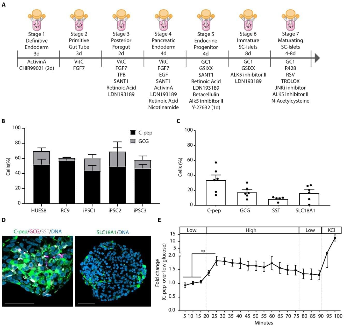
Stem cell-derived pancreatic islets are being studied as a rich transplantable source for insulin production, a therapeutic for type 1 diabetes that overcomes the need to obtain islet cells from deceased donors.

The first attempts to transplant to treat type 1 diabetes began half a century ago. Doctors then sought the pancreatic tissue of deceased donors from which islet-producing tissue was removed for transplants. The islets produce life-saving insulin. Substantial advancements and increased success rates have led to islet-cell transplants becoming an approved therapy in Canada and Europe. The technique is still considered investigational in the United States.

But in a series of new advances, a team of endocrinologists and regenerative medicine specialists in the Netherlands has developed methods that improve the production of stem cells used to generate insulin-making islets.

Prepare for pandemic NOW as new virus spreads to all 50 States

Leading health experts have warned that the US is staring down the barrel of another pandemic as bird flu spirals out of control on US farms.

So far, the H5N1 outbreak has affected nearly 1,000 dairy cow herds and resulted in more than 70 human cases, including the first confirmed death.

The US poultry industry is at significant risk, say experts from the Global Virus Network (GVN), particularly in areas with high-density farming and where personal protective practices may be lacking.

Aging will be cured within 20 years

Lately, there’s been growing pushback against the idea that AI will transform geroscience in the short term.
When Nobel laureate Demis Hassabis told 60 Minutes that AI could help cure every disease within 5–10 years, many in the longevity and biotech communities scoffed. Leading aging biologists called it wishful thinking — or outright fantasy.
They argue that we still lack crucial biological data to train AI models, and that experiments and clinical trials move too slowly to change the timeline.

Our guest in this episode, Professor Derya Unutmaz, knows these objections well. But he’s firmly on Team Hassabis.
In fact, Unutmaz goes even further. He says we won’t just cure diseases — we’ll solve aging itself within the next 20 years.

And best of all, he offers a surprisingly detailed, concrete explanation of how it will happen:
building virtual cells, modeling entire biological systems in silico, and dramatically accelerating drug discovery — powered by next-generation AI reasoning engines.

🧬 In this wide-ranging conversation, we also cover:

✅ Why biological complexity is no longer an unsolvable barrier.
✅ How digital twins could revolutionize diagnosis and treatment.
✅ Why clinical trials as we know them may soon collapse.
✅ The accelerating timeline toward longevity escape velocity.
✅ How reasoning AIs (like GPT-4o, o1, DeepSeek) are changing scientific research.
✅ Whether AI creativity challenges the idea that only biological minds can create.
✅ Why AI will force a new culture of leisure, curiosity, and human flourishing.
✅ The existential stress that will come as AI outperforms human expertise.
✅ Why “Don’t die” is no longer a joke — it’s real advice.

🎙️ Hosted — as always — by Peter Ottsjö (tech journalist and author of Evigt Ung) and Dr. Patrick Linden (philosopher and author of The Case Against Death).

/* */