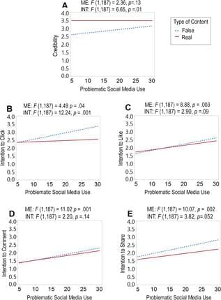
Toggle light / dark theme

For decades, researchers have sought ways to precisely manipulate and identify individual molecules like DNA in liquid environments. Such capabilities could revolutionize areas ranging from disease diagnosis to drug development. However, the randomness of molecular movements in fluids has hindered progress.

Now, scientists from Shenzhen University and the Chinese University of Hong Kong report promising advances in optical tweezing techniques that allow exquisite control over nanoscale biological particles (Light: Science & Applications, “CRISPR-powered optothermal nanotweezers: Diverse bio-nanoparticle manipulation and single nucleotide identification”).

A The diagrammatic sketch of the three components in the solution: DNA@AuNS conjugate, CRISPR/Cas12a complex, and target ssDNA. b Optical setup, the BS, SPF, and TL are beam splitter, short pass filter, and tube lens (f = 200 mm), respectively. Additional details of the setup are provided in the Materials and Methods section. c Dispersion of the three components in the solution without optical heating. d Optothermal net force induced migration and DNA@AuNS conjugate cleavage upon optical heating, the heating laser power is 0.5 mW. e Observation of the cleavage after the optical heating is switched off. (© Light: Science & Applications) (click on image to enlarge)

Pew Research Center surveys show that Americans are increasingly cautious about the growing role of AI in their lives generally. Today, 52% of Americans are more concerned than excited about AI in daily life, compared with just 10% who say they are more excited than concerned; 36% feel a mix of excitement and concern.

Despite these cautious overall views, Americans see some specific uses of AI positively, and attitudes depend a great deal on the context of how and why AI is being used.

This post summarizes what we know so far about how Americans view AI in everyday life, the workplace, and health and medicine.

While currently available PCSK9 inhibitor medications to lower cholesterol must be administered every few weeks by injection, a new, investigational PCSK9 inhibitor called recaticimab safely lowered bad cholesterol more than 50% when injected every 1–3 months, depending on dose.

A new PCSK9 inhibitor (recaticimab) injected every one to three months may work safely and provide more flexible dosing to lower cholesterol, according to late-breaking science presented today at the American Heart Association’s Scientific Sessions 2023. The meeting, Nov. 11–13, in Philadelphia, is a premier global exchange of the latest scientific advancements, research and evidence-based clinical practice updates in cardiovascular science.

“Previous studies found that 30% to 40% of people discontinued their current PCSK9 therapies, given every two to four weeks, during or after six months of beginning treatment. More flexible dosing with recaticimab, given up to every 12 weeks, might increase the proportion of people with high levels of bad cholesterol to stick with their recommended treatment to lower bad cholesterol levels and reduce risk of heart disease,” said lead study author Xin Du, Ph.D., a professor of cardiology at Beijing Anzhen Hospital and the Capital Medical University in Beijing, China.

Two Eötvös Loránd University researchers have made an exciting breakthrough in understanding how we age.

Researchers Dr. Ádám Sturm and Dr. Tibor Vellai from Eötvös Loránd University in Hungary have achieved a significant discovery in the study of aging. Their research centered on “transposable elements” (TEs) in our DNA

DNA, or deoxyribonucleic acid, is a molecule composed of two long strands of nucleotides that coil around each other to form a double helix. It is the hereditary material in humans and almost all other organisms that carries genetic instructions for development, functioning, growth, and reproduction. Nearly every cell in a person’s body has the same DNA. Most DNA is located in the cell nucleus (where it is called nuclear DNA), but a small amount of DNA can also be found in the mitochondria (where it is called mitochondrial DNA or mtDNA).

Meta, the parent company of Facebook, has made a groundbreaking development in brain-computer interface technology. They have unveiled an AI system that can decode visual representations and even “hear” what someone is hearing by studying their brainwaves. These advancements in brain-machine interface technology have the potential to transform our relationship with artificial intelligence and its potential applications in healthcare, communication, and virtual reality.

The University of Texas at Austin has developed a new technology that can translate brain activity into written text without surgical implants. This breakthrough uses functional Magnetic Resonance Imaging (fMRI) scan data to reconstruct speech. An AI-based decoder then creates text based on the patterns of neuronal activity that correspond to the intended meaning. This new technology could help people who have lost the ability to speak due to conditions such as stroke or motor neuron disease.

Despite the fMRI having a time lag, which makes tracking brain activity in real-time challenging, the decoder was still able to achieve impressive accuracy. The University of Texas researchers faced challenges in dealing with the inherent “noisiness” of brain signals picked up by sensors, but by employing advanced technology and machine learning, they successfully aligned representations of speech and brain activity. The decoder works at the level of ideas and semantics, providing the gist of thoughts rather than an exact word-for-word translation. This study marks a significant advance in non-invasive brain decoding, showcasing the potential for future applications in neuroscience and communication.

A speech prosthetic developed by a collaborative team of Duke neuroscientists, neurosurgeons, and engineers can translate a person’s brain signals into what they’re trying to say.

  • A pioneering speech prosthetic translates brain signals into speech, aiming to assist those with speech-affecting neurological disorders.
  • The device employs a high-density sensor array to capture brain activity with unprecedented detail.
  • New light activated cancer treatment.


    Several antibodies and antibody fragments have been previously developed for the treatment of various diseases, including cancer3,4. These antibodies bind to cell surface receptors expressed at higher levels on cancer cells, addressing a major challenge of selective cell targeting in cancer therapy. Although full-length antibodies have shown promise for treatment of several cancers, limited success has been demonstrated in eliminating solid tumors. Due to their large size, full-length antibodies are unable to diffuse deep into solid tumors5. In addition, it has been shown that high-affinity antibodies bind to the periphery of the tumor tissues, forming a barrier and preventing their further penetration6. Some studies in patients with cancer estimate that only 0.01% of the injected antibodies accumulate per gram of solid tumor tissue7. Small antibody fragments with low molecular weight can diffuse much deeper into tissues, presenting an excellent alternative to full-length antibodies. However, small antibody fragments have a low residence time in the body and often have a higher rate of dissociation (koff) from the target compared with full-length antibodies, limiting their clinical utility8. To address these challenges, antibody fragments are often multimerized9,10 and/or conjugated to larger proteins11, which increases the size of antibody fragments, again reducing their ability to penetrate into the tumor.

    One solution to overcome the limitation of low residence time would be to replace the noncovalent interactions between the antibody fragment and its antigen with a covalent bond. In a notable effort, an affibody containing a photocrosslinker in its antigen binding region was shown to covalently link to its antigen and demonstrated higher accumulation on tumor tissues12. Another pioneering study involved developing affibodies containing a latent bioreactive amino acid in their antigen binding region that forms a covalent bond with the target antigen by proximity-dependent reaction without any external impetus13. However, the former had substantially lower binding affinity compared with its wild-type (wt) counterpart and thus, requires using a high concentration for efficient initial binding, while the latter could react with target antigen expressed on healthy cells causing side effects.

    Although antibody-based therapeutics are more selective than several cytotoxic small molecule drugs used for cancer treatment, they can cause cardiac toxicity and skin reactions14. These side effects are due to the binding of the antibody to its receptor antigen expressed on healthy cells. This challenge could be addressed by activating antibody–antigen binding in the tumor microenvironment. One notable example in this direction is the development of antibodies containing an inhibitory N-terminal domain that is removed by tumor-specific proteases15. However, this approach would be difficult to extend to antibody fragments whose N terminus is not involved in antigen binding. We and others have also developed light-activated antibody fragments either by site-specific installation of photocaged functional groups or by introducing light-responsive proteins into antibodies16,17,18. In principle, such antibodies could be activated at the site of tumors using surgically implanted biocompatible light-emitting diodes (LEDs)19, thereby reducing the side effects of antibody-based therapeutics.

    This is a nontoxic version of cancer treatment that works on any type of cancer.


    Breast cancer is the most common cancer affecting women in Singapore. Treatment is multimodal and often involves surgery to remove the cancer and lymph nodes involved.

    Adjuvant therapy, given after the , is used to irradiate and destroy micrometastases, which are in the blood stream or lymphatics, to decrease recurrence. This form of therapy is subdivided into local (radiotherapy) and systemic therapy (endocrine therapy, chemotherapy and targeted therapy).

    Studies have shown that has increased with breast conserving therapy (BCT) where only the tumor and a margin is removed from the body post mastectomy, compared to full mastectomy alone, which removes all parts of the breast. For BCT, radiotherapy has to be administered after lumpectomy, which removes other from the breast and some normal tissue around it.

    Join us on Patreon! https://www.patreon.com/MichaelLustgartenPhD

    Discount Links:
    Epigenetic, Telomere Testing: https://trudiagnostic.com/?irclickid=U-s3Ii2r7xyIU-LSYLyQdQ6…M0&irgwc=1
    Use Code: CONQUERAGING

    NAD+ Quantification: https://www.jinfiniti.com/product/intracellular-nad-test/
    Use Code: ConquerAging At Checkout.

    Oral Microbiome: https://www.bristlehealth.com/?ref=michaellustgarten.
    Enter Code: ConquerAging.

    At-Home Metabolomics: https://www.iollo.com?ref=michael-lustgarten.
    Use Code: CONQUERAGING At Checkout.

    At-Home Blood Testing (SiPhox Health): https://getquantify.io/mlustgarten.