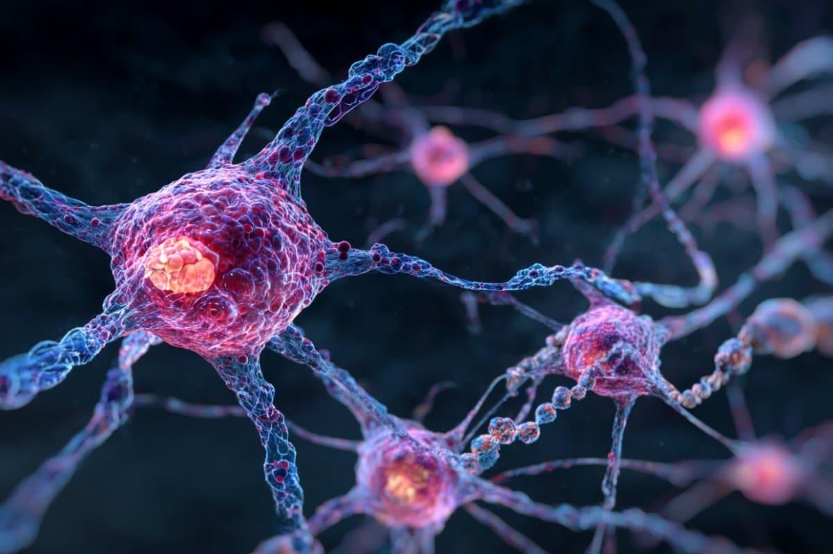
Kumar et al. show that under glucose-depleted conditions, neurons can use fatty acids as an alternative source of energy to support synaptic function.

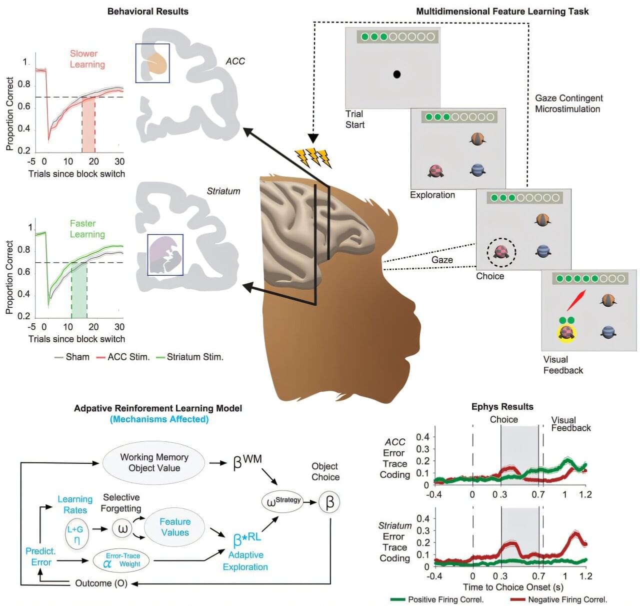
Research led by Thilo Womelsdorf, professor of psychology and biomedical engineering at the Vanderbilt Brain Institute, could revolutionize how brain-computer interfaces are used to treat disorders of memory and cognition.
The study, “Adaptive reinforcement learning is causally supported by anterior cingulate cortex and striatum,” was published June 10, 2025, in the journal Neuron.
According to researchers, neurologists use electrical brain-computer interfaces (BCIs) to help patients with Parkinson’s disease and spinal cord injuries when drugs and other rehabilitative interventions are not efficient. For these disorders, researchers say brain-computer interfaces have become electroceuticals that substitute pharmaceuticals by directly modulating dysfunctional brain signals.
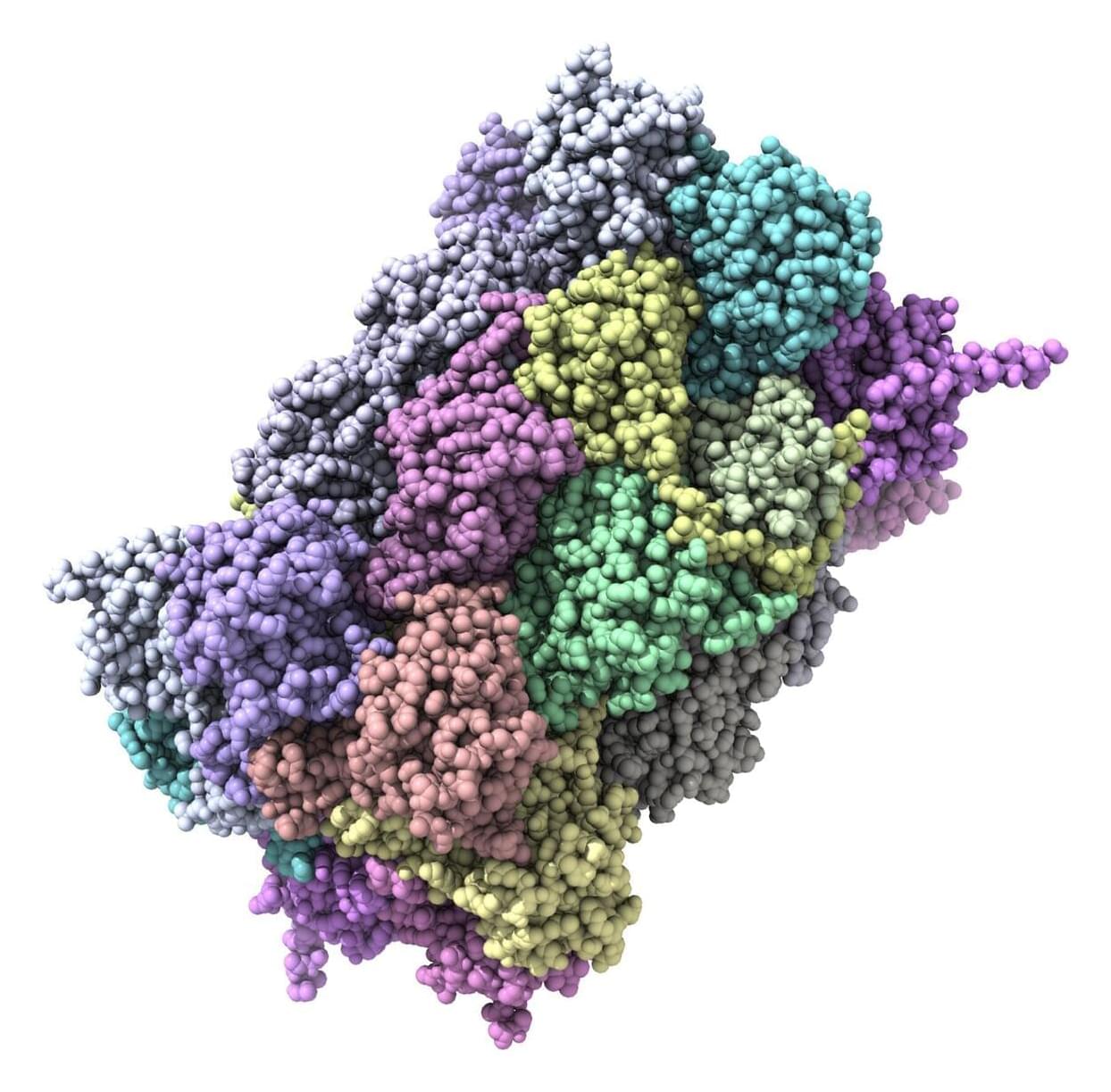
For decades, the story of Alzheimer’s research has been dominated by a battle between A-beta and tau amyloids, both of which can kill neurons and impact the brain’s ability to function. A new study suggests, however, that these sticky brain plaques may not be operating alone.
Johns Hopkins University researchers have identified more than 200 types of misfolded proteins in rats that could be associated with age-related cognitive decline.
The findings could lead the way to finding new therapeutic targets and treatments in humans that could provide relief for the millions of people over 65 who suffer from Alzheimer’s, dementia, or other diseases that rob them of their memories and independence as they age.
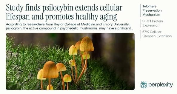
According to researchers from Baylor College of Medicine and Emory University, psilocybin, the active compound in psychedelic mushrooms, may have significant anti-aging properties, extending human cell lifespan by up to 57% in laboratory studies and improving survival rates in aged mice by 30% compared to untreated controls.
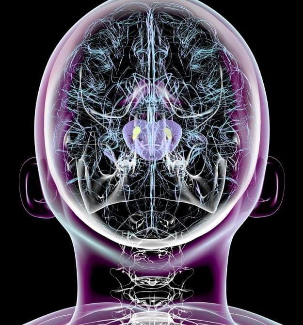
In 2017, he led a study that identified for the first time an abnormal form of a protein called SOD1 in Parkinson’s patients. Under normal conditions, this protein acts as an antioxidant enzyme, protecting brain cells from damage caused by free radicals, highly reactive molecules that contain oxygen and can deteriorate cells if not properly neutralized. Free radicals are produced by natural bodily processes as well as by external factors, like diet, smoking, and exposure to pollution.
In people with Parkinson’s disease, SOD1 suffers alterations that prevent it from fulfilling its protective function, with it instead accumulating in the brain and causing neuronal damage, according to the findings of Double’s team.
Based on these results, the team then conducted further research, with results suggesting that copper supplementation in the brain could be an effective way to slow and even reverse the symptoms of Parkinson’s (copper is crucial to SOD1’s function). To test this hypothesis, they evaluated the efficacy of a drug called CuATSM, designed to cross the blood-brain barrier and deliver copper directly to brain tissue.

From a brain chip that enabled a paralyzed patient to move his hand to a Pentagon-sponsored technology designed to restore memories, several exciting technologies have been announced recently that could advance the field of neurology. Here are 10 examples.
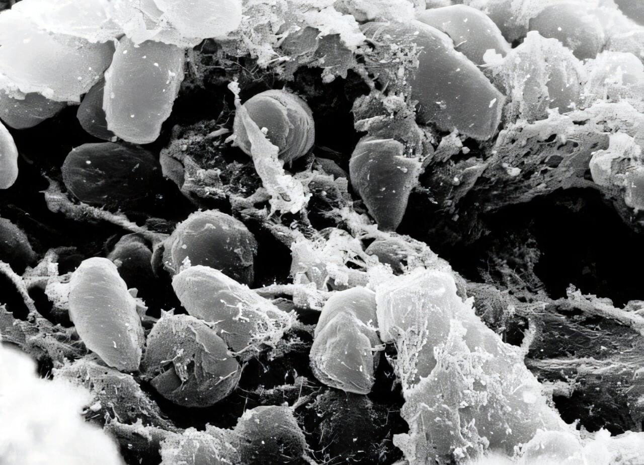
A research team led by Eske Willerslev, professor at the University of Copenhagen and the University of Cambridge, has recovered ancient DNA from 214 known human pathogens in prehistoric humans from Eurasia.
The study shows, among other things, that the earliest known evidence of zoonotic diseases—illnesses transmitted from animals to humans, like COVID in recent times—dates back to around 6,500 years ago, with such diseases becoming more widespread approximately 5,000 years ago.
“The spatiotemporal distribution of human pathogens in ancient Eurasia” is the largest study to date on the history of infectious diseases and has been published in Nature.
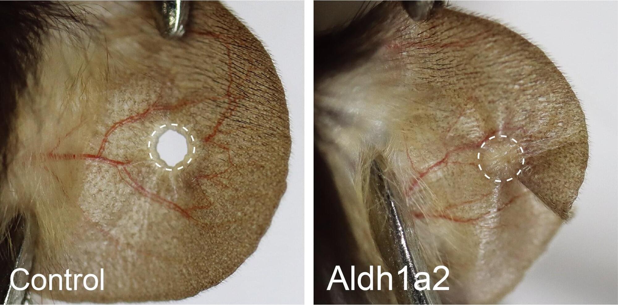
Research led by the National Institute of Biological Sciences in Beijing has discovered that switching on a single dormant gene enables mice to regenerate ear tissue.
Some vertebrates such as salamanders and fish can regenerate complex tissue structures with precision. A lost limb can be regrown, a damaged heart or eye can be repaired. Salamanders are so remarkable at reconstructing damaged tissues that even a spinal cord injury with severed neural motor connectivity can be restored.
Mammals occasionally showcase the ability to regenerate. Deer antlers and goat horns are examples of living tissue regeneration. Mice can regrow fingertips if they are lost. A healthy human liver can experience up to 70% loss of tissue and regrow to near full size within several weeks.