A groundbreaking study suggests that Parkinson’s disease may begin in the kidneys, where a toxic protein builds up and travels to the brain. This discovery could reshape our understanding of the disease’s origins and risk factors.

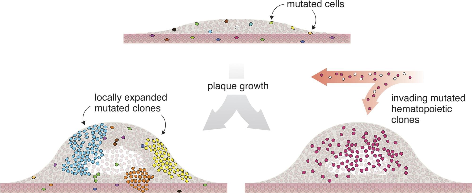
Researchers from the University of Southern Denmark and Odense University Hospital have studied tissue from patients with atherosclerosis. They found that many of the cells in the diseased tissue carried the same genetic alteration and appeared to originate from a single ancestral cell that had divided repeatedly—a pattern otherwise associated with tumor biology.
In several patients, a large proportion of the cells were derived from one single mutated cell that had undergone many rounds of cell division.
“It’s striking how many cells in the tissue share the exact same genetic change. In several samples, more than 10% of the cells—hundreds of thousands cells—carried the same alteration. It’s difficult to interpret this as anything other than all these cells originating from a shared ancestral cell that, at some point during disease development, acquired the mutation,” says Lasse Bach Steffensen, Associate Professor at the Department of Molecular Medicine at the University of Southern Denmark.

Every three seconds, someone in the world develops dementia. Alzheimer’s disease is the most common form of dementia, accounting for between 60% and 70% of all cases.
Although scientists have made significant progress in understanding the disease, there’s still no cure. That’s partly because Alzheimer’s disease has multiple causes—many of which are still not fully understood.
Two proteins which are widely believed to play central roles in Alzheimer’s disease are amyloid-beta and tau. Amyloid-beta forms sticky plaques on the outside of brain cells. This disrupts communication between neurons. Tau accumulates inside brain cells, where it twists into tangles. This ultimately leads to cell death. These plaques and tangles are the hallmark features of Alzheimer’s disease.
The human brain is made up of billions of interconnected cells that are constantly talking to each other. A new study published in Nature zooms in to the single-cell level to see how this cellular communication may be going wrong in brains affected by post-traumatic stress disorder (PTSD).
Until recently, researchers did not have the technology to study genetic variation within individual cells. But now that it’s available, a team led by Matthew Girgenti, Ph.D., assistant professor of psychiatry at Yale School of Medicine, has been analyzing brain cells to uncover genetic variants that might be associated with psychiatric diseases such as major depressive disorder (MDD) and PTSD.
Their latest study is one of the first to examine a major psychiatric disorder, PTSD, at the single-cell level. For years, doctors have been prescribing antidepressants to treat the condition because there are currently no drugs specifically designed for PTSD. Girgenti hopes that identifying novel molecular signatures associated with the psychiatric disease can help researchers learn how to develop new drugs or repurpose existing ones to treat it more effectively.
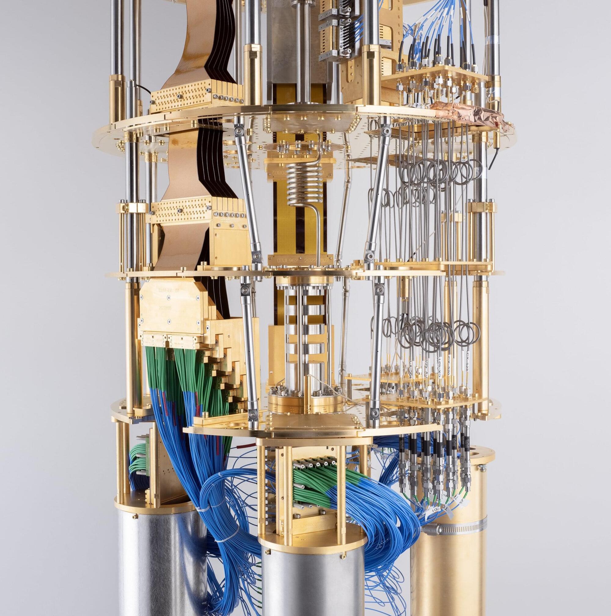
Quantum computers have the potential to speed up computation, help design new medicines, break codes, and discover exotic new materials—but that’s only when they are truly functional.
One key thing that gets in the way: noise or the errors that are produced during computations on a quantum machine—which in fact makes them less powerful than classical computers —until recently.
Daniel Lidar, holder of the Viterbi Professorship in Engineering and Professor of Electrical & Computer Engineering at the USC Viterbi School of Engineering, has been iterating on quantum error correction, and in a new study along with collaborators at USC and Johns Hopkins, has been able to demonstrate a quantum exponential scaling advantage, using two 127-qubit IBM Quantum Eagle processor-powered quantum computers, over the cloud.
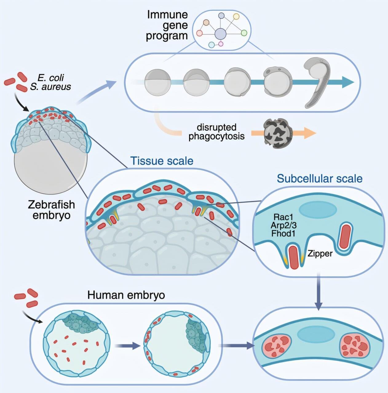
Research led by scientists from the Institute of Molecular Biology of Barcelona (IBMB) of the CSIC and the Bellvitge Biomedical Research Institute (IDIBELL) has managed to film how a few days-old embryos defend themselves from a potential infection by bacteria. The work is published this week in the journal Cell Host and Microbe.
Specifically, they have been able to see how zebrafish embryos use cells present on their surface, known as epithelial cells, to ingest and destroy bacteria through a process called phagocytosis, similar to that carried out by white blood cells. Crucially, scientists could observe that this ability to eliminate bacteria is also present in human embryos.
Using state-of-the-art microscopy techniques, the research shows how cells capture Escherichia coli and Staphylococcus aureus bacteria through small protrusions of their membrane, in which the protein Actin is involved. “Our research shows that, at the beginning of development—before implantation in the uterus and before the formation of organs—embryos already have a defense system that allows them to eliminate bacterial infections,” says Esteban Hoijman, researcher at IBMB-CSIC and IDIBELL, leader of the research.
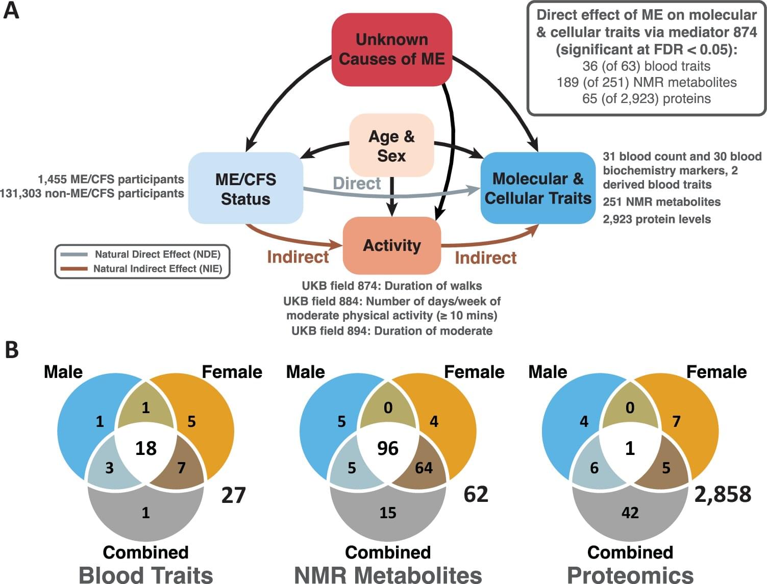
People with ME/CFS (myalgic encephalomyelitis/chronic fatigue syndrome) have significant differences in their blood compared with healthy individuals, a new study reveals, suggesting a path toward more reliable diagnosis of the long-term debilitating illness. The paper is published in the journal EMBO Molecular Medicine.
The largest ever biological study of ME/CFS has identified consistent blood differences associated with chronic inflammation, insulin resistance, and liver disease.
Significantly, the results were mostly unaffected by patients’ activity levels, as low activity levels can sometimes hide the biological signs of illness, experts say.