Toggle light / dark theme

Childhood stress strongly linked to chronic disease in adulthood, researchers report

Research published by Duke University researchers has found a strong link between higher stress in children and adverse health conditions for them later in life. Appearing in the journal Proceedings of the National Academy of Sciences, the study used measurable metrics of health over time to create a more quantitative view of how stress early in life affects health.

“We’ve had an idea for a long time, since the ’80s at least, that when children have adversity in their lives, it affects how their bodies work, not just psychologically, but also physiologically. It gets underneath the skin, and it becomes embodied in the way your body handles stress,” said co-author Herman Pontzer, Duke professor of evolutionary anthropology and global health.

Researchers focused on (AL), which refers to the wear and tear on the body because of chronic stress. The researchers “tested associations between childhood AL and adult cardiometabolic health,” relying on biomarkers that included antibodies of C-reactive protein, which is a marker of inflammation in the body; and the Epstein-Barr virus, which is common and highly contagious; body mass index; and .

New protein interaction map sheds light on how brain cell communication breaks down in Alzheimer’s disease

A new study led by the Icahn School of Medicine at Mount Sinai offers one of the most comprehensive views yet of how brain cells interact in Alzheimer’s disease, mapping protein networks that reveal communication failures and point to new therapeutic opportunities.

Published online in Cell, the study analyzed in brain tissue from nearly 200 individuals.

The researchers discovered that disruptions in communication between neurons and supporting called glia—specifically astrocytes and microglia—are closely linked to the progression of Alzheimer’s disease. One protein in particular, called AHNAK, was identified as a top driver of these harmful interactions.

Theoretical model uses neuroimaging data to link brain alterations to schizophrenia

Schizophrenia is a chronic mental health disorder characterized by hallucinations, delusions, disorganized thinking and atypical movement or speech patterns. This psychiatric condition can be highly debilitating, and diagnosed individuals can report markedly different experiences.

Understanding the neurobiological basis of could be highly valuable, as it could inform the development of new interventions to reduce the risk of its emergence or treat its symptoms. The results of many neuroimaging studies carried out so far, however, were inconsistent or inconclusive, failing to clearly delineate the processes and brain regions implicated in its clinical expression.

In a recent paper published in Nature Mental Health, researchers at Taipei Medical University analyzed meta-analyses summarizing the most consistent findings of schizophrenia-related neuroimaging studies. Drawing on the results of this analysis, they developed a new theoretical model that delineates characteristic brain alterations linked to the psychiatric disorder.

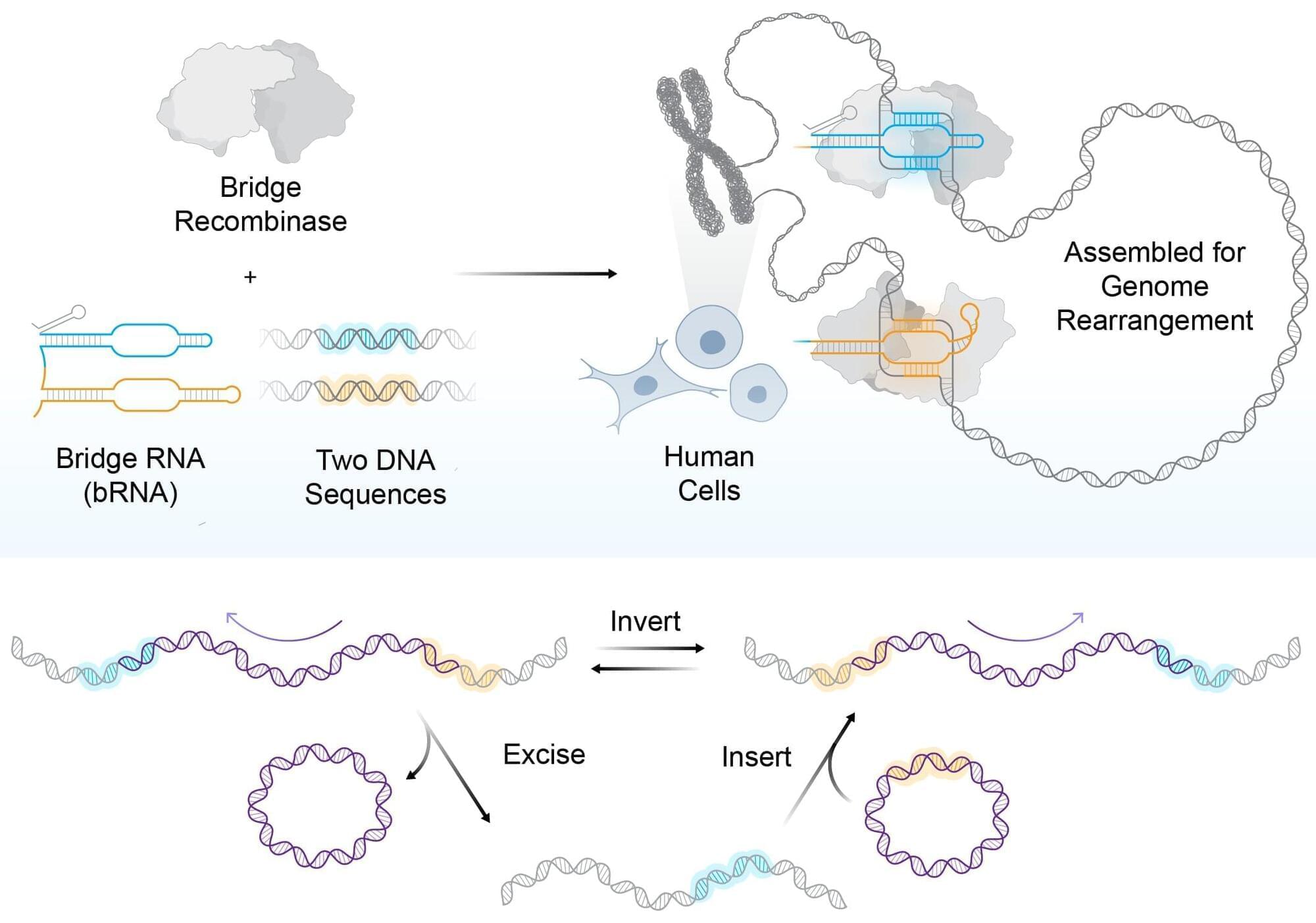
Bridge recombinases, optimized for human cells, enable massive programmable DNA rearrangements

For decades, gene-editing science has been limited to making small, precise edits to human DNA, akin to correcting typos in the genetic code. Arc Institute researchers are changing that paradigm with a universal gene editing system that allows for cutting and pasting of entire genomic paragraphs, rearranging whole chapters, and even restructuring entire passages of the genomic manuscript.

AI system for rapid annotation of medical images could accelerate clinical research

Annotating regions of interest in medical images, a process known as segmentation, is often one of the first steps clinical researchers take when running a new study involving biomedical images.

For instance, to determine how the size of the brain’s hippocampus changes as patients age, the scientist first outlines each hippocampus in a series of brain scans. For many structures and image types, this is often a manual process that can be extremely time-consuming, especially if the regions being studied are challenging to delineate.

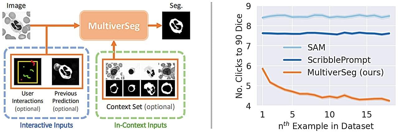
To streamline the process, MIT researchers developed an artificial intelligence-based system that enables a researcher to rapidly segment new biomedical imaging datasets by clicking, scribbling, and drawing boxes on the images. This new AI model uses these interactions to predict the segmentation.

/* */