Stanford Medicine researchers have developed an artificial intelligence tool to help scientists better plan gene-editing experiments.


Scientists said Wednesday that they had created an AI model able to predict medical diagnoses years in advance, building on the same technology behind consumer chatbots like ChatGPT.
Based on a patient’s case history, the Delphi-2M AI “predicts the rates of more than 1,000 diseases” years into the future, the team from British, Danish, German and Swiss institutions wrote in a paper published in the journal Nature.
Researchers trained the model on data from Britain’s UK Biobank – a large-scale biomedical research database with details on about half a million participants.

University of Oxford-led research finds low-dose rapamycin functions as a genomic protector in aging human immune cells, lowering DNA damage.
The mechanistic target of rapamycin (mTOR) is a central signaling pathway that regulates and coordinates cell growth, metabolism, and survival in response to environmental cues. It helps cells integrate signals from growth factors, nutrients, and stress to control whether they are in an anabolic (building up) or catabolic (breaking down) state.
Aging immune systems accumulate DNA damage linked to immunosenescence. Rapamycin is a drug that inhibits the mTOR pathway. Originally developed for organ transplantation to prevent immune rejection, previous research has found that, at non-immunosuppressive doses, rapamycin can mitigate cellular senescence.
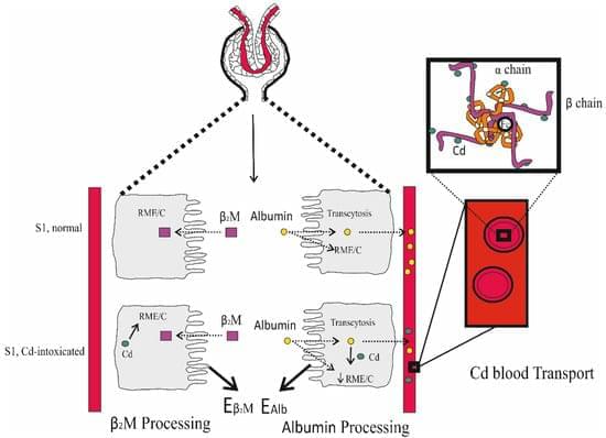
Cadmium (Cd) is a metal with no nutritional value or physiological role. However, it is found in the body of most people because it is a contaminant of nearly all food types and is readily absorbed. The body burden of Cd is determined principally by its intestinal absorption rate as there is no mechanism for its elimination. Most acquired Cd accumulates within the kidney tubular cells, where its levels increase through to the age of 50 years but decline thereafter due to its release into the urine as the injured tubular cells die. This is associated with progressive kidney disease, which is signified by a sustained decline in the estimated glomerular filtration rate (eGFR) and albuminuria. Generally, reductions in eGFR after Cd exposure are irreversible, and are likely to decline further towards kidney failure if exposure persists.

Scientists have developed and tested a deep-learning model that could support clinicians by providing accurate results and clear, explainable insights—including a model-estimated probability score for autism.
The model, outlined in a study published in eClinicalMedicine, was used to analyze resting-state fMRI data—a non-invasive method that indirectly reflects brain activity via blood-oxygenation changes.
In doing so, the model achieved up to 98% cross-validated accuracy for Autism Spectrum Disorder (ASD) and neurotypical classification and produced clear, explainable maps of the brain regions most influential to its decisions.
Researchers led by Rice University’s Yong Lin Kong have developed a soft but strong metamaterial that can be controlled remotely to rapidly transform its size and shape.
The invention, published in Science Advances, represents a significant advancement that can potentially transform ingestible and implantable medical devices.
Metamaterials are synthetic constructs that exhibit unusual properties not typically found in natural materials. Instead of relying solely on chemical composition, the effective behavior of these materials is primarily determined by the physical structure, i.e., the specific shape, arrangement and scale of their building blocks.

The brain is famously plastic: Neurons’ ability to change their behavior in response to new stimuli is what makes learning possible. And even neurons’ response to the same stimuli changes over time—a phenomenon known as representational drift. Yet our day-to-day perception of the world is relatively stable. How so?
Resolving such puzzles matters for future brain-computer interfaces, sensory prostheses and therapies for neurological disease. On a quest for an answer, Rice University scientists have built ultraflexible probes thousands of times thinner than a human hair and used them to track neurons in the visual cortex of mice for 15 consecutive days as the animals viewed thousands of images—from line patterns to pictures of the natural world.
The devices, called nanoelectronic threads (NETs), embed seamlessly with brain tissue, allowing for high-fidelity chronic recordings of brain activity.