When it comes to their survival, cancer cells have a host of backup plans.
This is especially true of the nutrients that cancers use to grow and spread. In addition to relying on sugars like glucose to power their proliferation, some cancer cells also use ketones — metabolites produced from fats when the body is fasting or on a low carb diet — as an alternate fuel source.
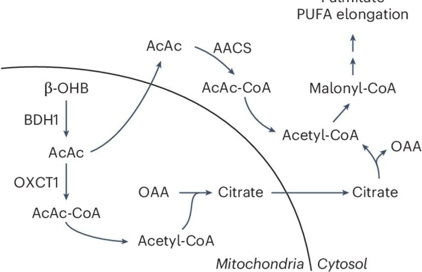
Now, a new study scientists suggest that the routes cancer cells use to process these different nutrients deeply influence cell behavior. They discovered an alternate, or non-canonical, path by which cancer cells convert a ketone called β-hydroxybutyrate (β-OHB) into acetyl-CoA, an essential metabolic building block for fatty acids and cholesterol that supports cell proliferation.
The findings, published in the journal Nature Metabolism, could reshape how the relationship between diet and cancer is viewed.
The authors also found that cancer cells can leverage this alternative β-OHB pathway even when glucose, the body’s main source of energy, is plentiful. This suggests that, depending on the circumstances, glucose may not always be the nutrient of choice for cells.


