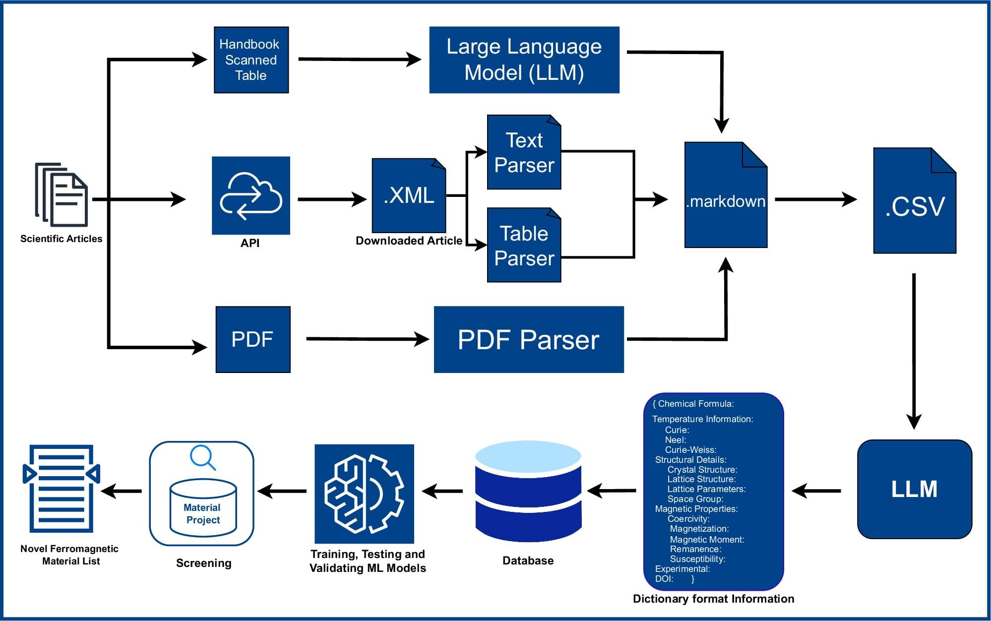
Toggle light / dark theme

New Strategy Enhances CRISPR Editing Efficiency for Therapeutic Use

While CRISPR-mediated gene editing has led to powerful advances across biology, medicine, and agriculture, challenges persist in optimizing the editing efficiency of enzymes, such as the widely used Cas9 nuclease. This is especially true in therapeutic use cases, where the goal is to attain high rates of editing via a relatively low and transient enzyme dose.

In a new study published in the April 2025 issue of The CRISPR Journal titled, “Hairpin Internal Nuclear Localization Signals in CRISPR-Cas9 Enhance Editing in Primary Human Lymphocytes,” researchers from the Innovative Genomics Institute (IGI) at the University of California (UC), Berkeley, present a strategy to improve editing efficiency in human immune cells for therapeutic applications by leveraging new constructs for nuclear localization signal (NLS) sequences.

“Efficient CRISPR enzyme production is essential for translation. This is one element that allowed the rapid clinical evaluation of Casgevy, the world’s first genome editing drug. Unfortunately, this aspect tends to be overlooked in the basic research performed in academia,” said Ross Wilson, PhD, assistant adjunct professor of molecular and cell biology at UC Berkeley, who led the new study.

Delineating inflammatory from non-inflammatory mechanisms for therapy optimization in psoriatic arthritis

In this Perspective, the authors propose that patients with psoriatic arthritis and an inadequate response to therapy can be classified into two distinct subgroups, characterized by persistent inflammatory and non-inflammatory phenotypes, and discuss potential mechanisms underlying these phenotypes, as well as considerations for treatment strategies and trial design.

Scientists reveal cilia’s secrets using connectome data

Many cells in our body have a single primary cilium, a micrometer-long, hair-like organelle protruding from the cell surface that transmits cellular signals. Cilia are important for regulating cellular processes, but because of their small size and number, it has been difficult for scientists to explore cilia in brain cells with traditional techniques, leaving their organization and function unclear.

In a new series of work, researchers at HHMI’s Janelia Research Campus, the Allen Institute, the University of Texas Southwestern Medical Center, and Harvard Medical School used super high-resolution 3D electron microscopy images of mouse brain tissue generated for creating connectomes to get the best look yet at primary cilia.

Vast expands Haven-1 Lab partnerships with biotech and space science leaders

Non-personalized content and ads are influenced by things like the content you’re currently viewing and your location (ad serving is based on general location). Personalized content and ads can also include things like video recommendations, a customized YouTube homepage, and tailored ads based on past activity, like the videos you watch and the things you search for on YouTube. We also use cookies and data to tailor the experience to be age-appropriate, if relevant.

Select “More options” to see additional information, including details about managing your privacy settings. You can also visit g.co/privacytools at any time.

Neuroscientists develop AI tool to unlock cerebellum’s secrets

Understanding and treating brain disorders such as tremor, imbalance, and speech impairments requires deep knowledge of the cerebellum, a part of the brain that’s crucial for making accurate movements.

Scientists have long been able to eavesdrop on and record the electrical signals transmitted by neurons () in the cerebellum, allowing them to observe the signals entering and exiting this region. But the computations that the brain performs between the input and output have been largely a mystery.

However, that is now changing. A team of researchers, including those from Baylor College of Medicine, have created an artificial intelligence tool that can identify the type of neuron producing electrical signals recorded from the cerebellum during behavior, allowing a new understanding of how the cerebellum works.

/* */