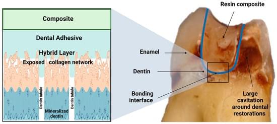
Damage in the bonding interface is a significant factor that leads to premature failure of dental bonded restorations. The imperfectly bonded dentin-adhesive interface is susceptible to hydrolytic degradation and bacterial and enzyme attack, severely jeopardizing restorations’ longevity. Developing caries around previously made restorations, also called “recurrent or secondary caries,” is a significant health problem. The replacement of restorations is the most prevailing treatment in dental clinics, leading to the so-called “tooth death spiral”. In other words, every time a restoration is replaced, more tooth tissue is removed, increasing the size of the restorations until the tooth is eventually lost. This process leads to high financial costs and detriment to patients’ quality of life.
Not every part of a DNA sequence gets translated into a protein. Each sequence consists of exons, which are included in the final RNA transcript, and introns, which are thrown away.
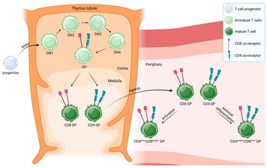
The expression of CD4 and CD8 co-receptors defines two distinct T cell populations with specialized functions. While CD4+ T cells support and modulate immune responses through different T-helper (Th) and regulatory subtypes, CD8+ T cells eliminate cells that might threaten the organism, for example, virus-infected or tumor cells. However, a paradoxical population of CD4+CD8+ double-positive (DP) T cells challenging this paradigm has been found in the peripheral blood. This subset has been observed in healthy as well as pathological conditions, suggesting unique and well-defined functions. Furthermore, DP T cells express activation markers and exhibit memory-like features, displaying an effector memory (EM) and central memory (CM) phenotype.
A series of focused interviews with the most interesting and impactful thought leaders in the field. We press on first principles—how they define core ideas in their domain, how they see the present, and where they believe intelligence is headed. Brought to you by SingularityNET and the AGI Society.
About this interview — Joscha Bach. In this kickoff episode, cognitive scientist Joscha Bach explores consciousness as a coherence-forming learning process, argues for a computational view of mind, and outlines why machine consciousness should be treated as a testable hypothesis rather than a slogan. He discusses the California Institute for Machine Consciousness, contrasts today’s “idiot-savant” AI with developmental intelligence, sketches futures from universal basic intelligence to post-human infospheres, and offers frank advice to new researchers on pursuing bold, technically grounded work.
SingularityNET was founded by Dr. Ben Goertzel with the mission of creating a decentralized, democratic, inclusive, and beneficial Artificial General Intelligence (AGI). An AGI is not dependent on any central entity, is open to anyone, and is not restricted to the narrow goals of a single corporation or even a single country.
The SingularityNET team includes seasoned engineers, scientists, researchers, entrepreneurs, and marketers. Our core platform and AI teams are further complemented by specialized teams devoted to application areas such as finance, robotics, biomedical AI, media, arts, and entertainment.
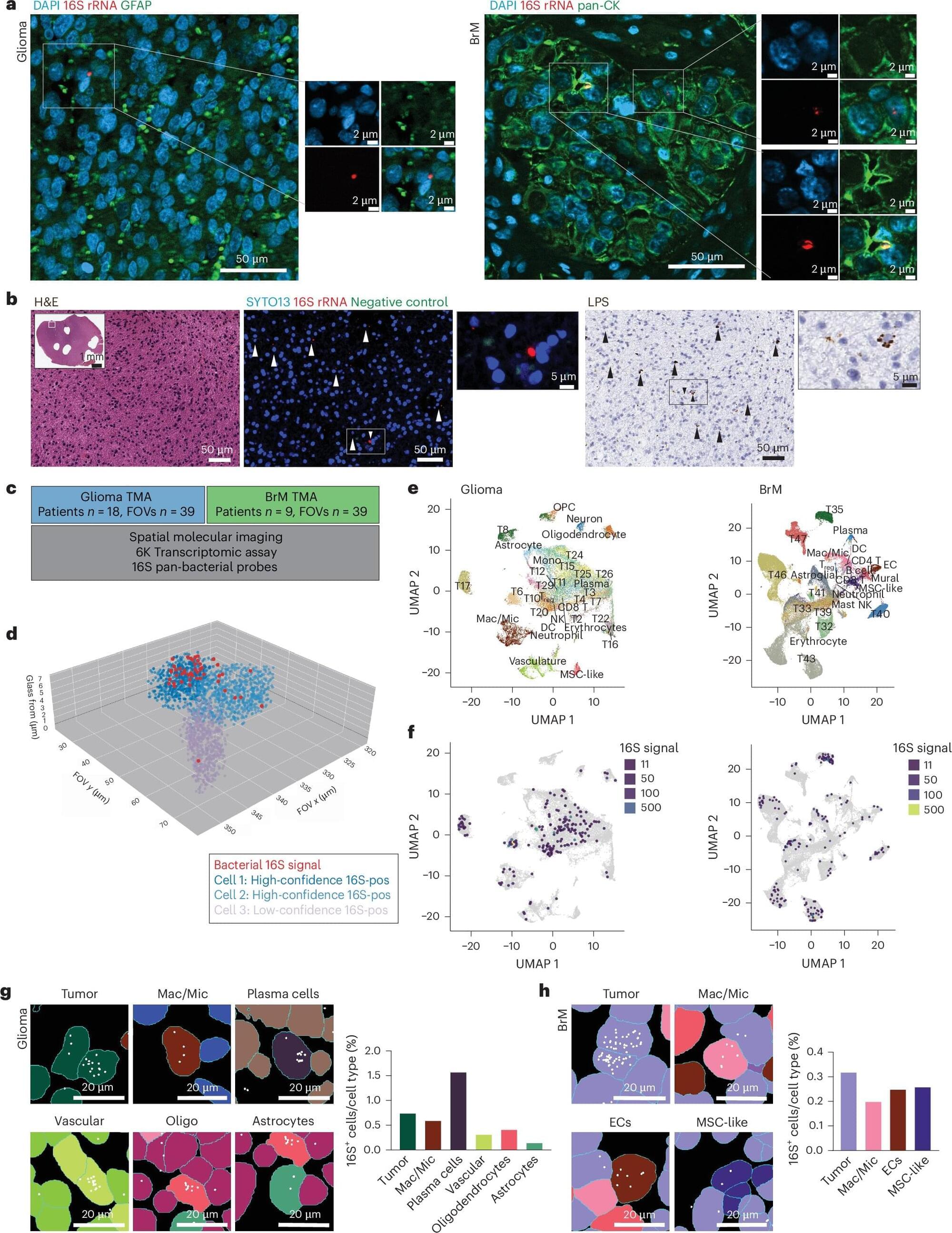
Researchers at The University of Texas MD Anderson Cancer Center have uncovered unexpected traces of bacteria within brain tumors. This discovery offers new insights into the environment in which brain tumors grow and sets the stage for future studies seeking to improve treatment outcomes.
Published today in Nature Medicine, the data revealed that bacterial genetic and cellular elements were present inside brain tumor cells and across the tumor microenvironment. These bacterial components appeared biologically active, potentially influencing tumor behavior and progression in patients with gliomas and brain metastases.
The multi-institutional study was led by Golnaz Morad, D.D.S, Ph.D., postdoctoral research fellow in Surgical Oncology, and Jennifer Wargo, M.D., professor of Surgical Oncology and Genomic Medicine and core member of the James P. Allison Institute—working in close collaboration with MD Anderson’s Platform for Innovative Microbiome and Translational Research (PRIME-TR).
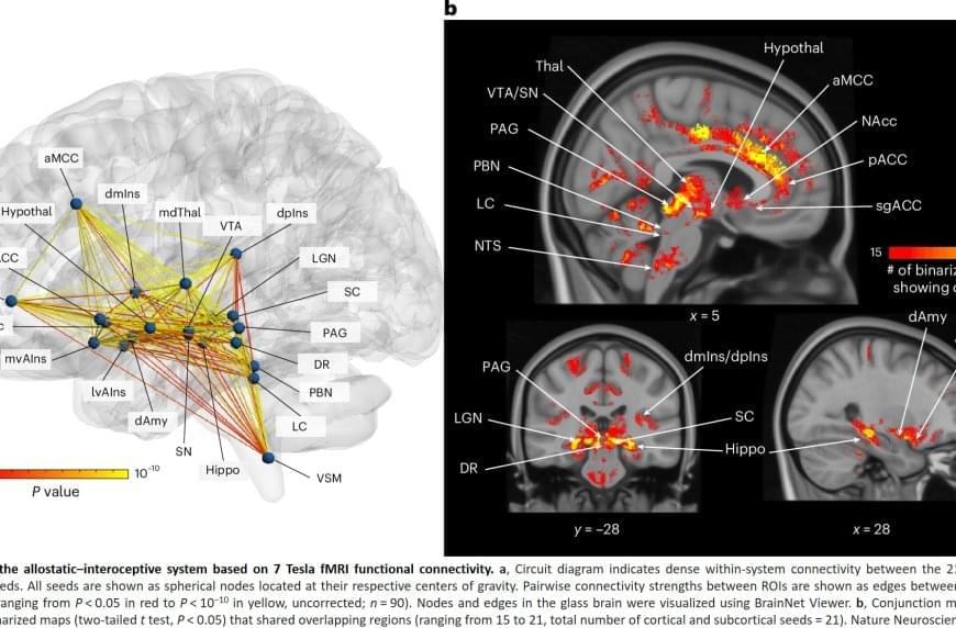
They also used a recently validated map of deep brain areas. This in vivo atlas, Brainstem Navigator, maps the regions involved in regulating the autonomic, immune and endocrine systems.
The authors analytic approach was guided by decades of basic research that has identified two main brain pathways in mammals: one set of pathways (allostatic) that sends signals from the brain to control the body’s organs, and the other set (interoceptive) that sends signals from the body to the brain, informing it about what’s happening inside us.
The findings replicated and expanded on their previous 3 Tesla work, confirming nearly all the direct connections identified in non-human mammals: 100% of those between cortical areas and 96% of those linking subcortical areas to both cortical and other subcortical areas. As expected, the authors found two-way connections between the brain areas that help manage the body’s needs (like the anterior cingulate cortex) and the areas that sense what’s happening inside the body (like the posterior insula). This means these regions communicate back and forth, helping the brain predict and regulate what the body needs.
Mounting evidence suggests that one of the brain’s central roles is to anticipate and meet the body’s energy needs. The findings place the monitoring and regulation of the body’s needs at the functional core of the human brain, showing the close connection between mental and physical health.
Previous studies in both animal models and humans have pointed to the existence of a distributed system in the brain that helps it anticipate and prepare for the body’s energy needs — a process called allostasis — as well as monitor the sensory conditions inside the body, known as interoception.
In an earlier study using 3 Tesla fMRI, the team mapped a network supporting allostasis and interoception in the human brain, but the comparatively limited spatial resolution and sensitivity of the 3 Tesla technology made it difficult to fully capture the system’s smaller structures in the brainstem, which are known to play a key role in these processes.
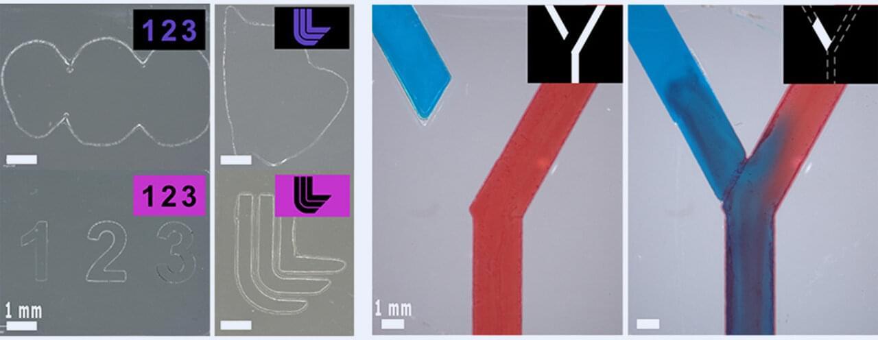
Additive manufacturing, or 3D printing, is normally a one-way street. In a digital light processing (DLP) printer, a structured pattern is projected onto a layer of liquid resin, which cures and solidifies. This builds an object up, layer-by-layer. But if the print isn’t exactly right, there’s no easy way to fix it after the fact: it usually ends up in the trash.
In a new study, published in Advanced Materials Technologies, researchers at Lawrence Livermore National Laboratory (LLNL) developed a hybrid additive and subtractive manufacturing system with a unique resin that enhances traditional 3D printing by introducing dual-wavelength behavior. Under blue light, the resin cures and hardens. Under ultraviolet light, it degrades back into a liquid. The hybrid printing system enables corrective manufacturing, provides improved print resolution and allows for upcycling and recycling of parts.
“Imagine if a company needed a part to fit a certain machine but it’s a prototype and they’re not quite sure what they want,” said LLNL scientist and author Benjamin Alameda. “They could theoretically print with our resin. And if there were defects or something they wanted to change about it, they don’t have to print a whole new part. They could just shine another wavelength on it and modify the existing part. That’s useful and less wasteful.”
Cells have an internal skeleton that maintains their structure and also drives their movement. Known as the cytoskeleton, this scaffold is composed of a network of dynamic filaments made of a protein called actin.
Given how important these structures are, alterations in the proteins that work together to build and control the actin cytoskeleton are often lethal or cause severe effects. For example, children born with mutations in the ARPC5 protein, which is part of the Arp2/3 complex, experience immunodeficiency and a high risk of fatal sepsis in early life.
“This is a rare and devastating condition, and until recently, it wasn’t clear how these mutations lead to such severe illness,” says Michael Way, who runs the Cellular Signaling and Cytoskeletal Function Laboratory at the Crick. “The only known effective treatment would involve early bone marrow transplantation to replace the faulty immune cells with ones which have a healthy actin cytoskeleton.”
After numerous successful trials in the model, the team sought to demonstrate what the microrobot could achieve under real clinical conditions. First, they were able to demonstrate in pigs that all three navigation methods worked and that the microrobot remains clearly visible throughout the entire procedure. The investigators then navigated microrobots through the cerebral fluid of a sheep.
“This complex anatomical environment has enormous potential for further therapeutic interventions, which is why we were so excited that the microrobot was able to find its way in this environment too,” Landers noted. “In vivo experiments conducted with an ovine model demonstrated the platform’s ability to operate within anatomically constrained regions of the central nervous system,” the investigators stated in their paper. “Furthermore, in a porcine model, all locomotion strategies were validated under clinical conditions, confirming precise microrobot navigation within the cerebrovascular system and highlighting the system’s compatibility with versatile in vivo environments.”
In addition to treating thrombosis, these new microrobots could also be used for localized infections or tumors. At every stage of development, the research team has remained focused on their goal, which is to ensure that everything they create is ready for use in operating theaters as soon as possible. The next goal is to look at human clinical trials. “The use of materials that have been FDA approved for other intravascular applications, coupled with the modular design of the robotic platform, should simplify translation and adaptability to a range of clinical workflows,” the authors concluded. Speaking about what motivates the whole team, Landers said, “Doctors are already doing an incredible job in hospitals. What drives us is the knowledge that we have a technology that enables us to help patients faster and more effectively and to give them new hope through innovative therapies.”