Toggle light / dark theme

Scientists are embarking on a £1.1 million project aimed at revolutionising drug production by using food by-products to develop new antimicrobial drugs.

Led by the University of Strathclyde in collaboration with the University of Surrey and GSK, the research endeavours to make antimicrobial production more cost-effective and sustainable, thereby addressing the pressing global challenge of antimicrobial resistance.

The project seeks to leverage bacteria, particularly Streptomyces, known for their potential to produce various drugs including antimicrobials. By harnessing food by-products, the team is aiming to device a less carbon-intensive process for biomanufacturing, which could pave the way for a range of medications including anti-parasitic, anti-cancer, anti-fungal, and immunosuppressant drugs.

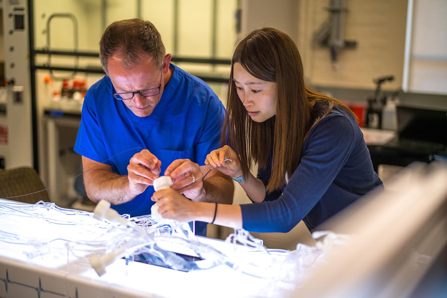
Researchers in the UC Davis Department of Biomedical Engineering are introducing a groundbreaking catheter-based device that could revolutionize heart attack and stroke prevention by enhancing intravascular imaging of dangerous plaques.


Researchers in the Department of Biomedical Engineering at the University of California, Davis, have developed a new catheter-based device that combines two powerful optical techniques to image the dangerous plaques that can build up inside the arteries that supply blood to the heart. By providing new details about plaque, the device could help clinicians and researchers improve treatments for preventing heart attacks and strokes.

Atherosclerosis occurs when fats, cholesterol and other substances accumulate on the artery walls, which can cause these vessels to become thick and stiff. A heart attack or stroke may occur if an atherosclerotic plaque inside the blood vessels ruptures or parts of it break off.

“Atherosclerosis, leading to heart attacks and strokes, is the number one cause of death in Western societies — exceeding all combined cancer types — and, therefore, a major public health issue,” said Laura Marcu, research team member leader and professor of biomedical engineering at UC Davis. “Better clinical management made possible by advanced intravascular imaging tools will benefit patients by providing more accurate information to help cardiologists tailor treatment or by supporting the development of new therapies.”

Space travel is associated with an increased incidence of headaches among astronauts, emphasizing the need for further research and improved treatment methods.

Space travel and zero gravity can take a toll on the body. A new study has found that astronauts with no prior history of headaches may experience migraine and tension-type headaches during long-haul space flight, which includes more than 10 days in space. The study was published in the March 13, 2024, online issue of Neurology, the medical journal of the American Academy of Neurology.

Understanding Space-Induced Headaches

Cellulose is the primary component of the cell walls of plants, making it the most common polymer on Earth. It’s responsible for the properties of materials like wood and cotton and is the primary component of dietary fiber, so it’s hard to overstate its importance to humanity.

Given its ubiquity and the fact that it’s composed of a bunch of sugar molecules linked together, its toughness makes it very difficult to use as a food source. The animals that manage to extract significant calories from cellulose typically do so via specialized digestive tracts that provide a home for symbiotic bacteria—think of the extra stomachs of cows and other ruminants.

Amazingly, humans also play host to bacteria that can break down cellulose—something that wasn’t confirmed until 2003 (long after I’d wrapped up my education). Now, a new study indicates that we’re host to a mix of cellulose-eating bacteria, some via our primate ancestry, and others through our domestication of herbivores such as cows. But urban living has caused the number of these bacteria to shrink dramatically.

A soft brain implant that unfurls under the skull could give doctors a less invasive way to monitor patients’ brain activity — and maybe allow people to directly control technology with their minds.

The challenge: Placing an electrode array on the surface of the brain allows scientists to see neural activity in far more detail than is possible with electrodes outside of the skull.

Known as “electrocorticography,” this technology was initially developed to treat people with epilepsy, but it’s also been used in research for other types of neurological disorders and in the development of brain-computer interfaces.

Denmark: A recent study published in JAMA Network Open has suggested an association between tooth agenesis and specific cancer types, specifically in childhood and early adulthood.

“Our population-based cohort study of 2.5 million live-born singletons with up to 40 years of follow-up showed that tooth agenesis was positively associated with several cancer types, including nephroblastoma, neuroblastoma, and hepatoblastoma in childhood; osteosarcoma in adolescence; and carcinomas of the bladder and colorectal carcinomas in young adulthood,” the researchers reported.

Previous studies have provided some evidence that tooth agenesis (congenital absence of one or more teeth) is associated with cancer risk, particularly carcinomas of the ovaries and colon, but these results are conflicting, and associations have not been evaluated yet in a population-based setting. To fill this knowledge gap, Saga Elise Eiset, Department of Clinical Medicine, Aarhus University, Aarhus, Denmark, and colleagues aimed to investigate the association between tooth agenesis and specific cancer types before 40 years of age.

Diabetic medication has garnered popularity within the last few years. It is not necessarily the need for these medications that is associated with its popularity, but a side effect that most individuals desire: weight loss. Since the advent of drugs that promote rapid weight loss such as Ozempic, Mounjaro, and Wegovy, widespread safety concerns are being raised regarding long-term effects and more immediate risks such as complications during surgery.

Ozempic, Mounjaro, and Wegovy are all part of a class of drugs known as semaglutides, which are glucagon-like peptide-1 receptor agonists or GLP1-RAs. GLP1 receptors are expressed on different cells within tissues and organs including the pancreas. GLP1-RAs help the pancreas release insulin and lower blood sugar levels, which makes these medications very helpful for those with diabetes. However, the stomach also has GLP1 receptors. Consequently, it activates these receptors as well and causes the stomach to digest food at a much slower rate. This delayed gastric emptying results in patients feeling full and not eating as much to lose weight. Additionally, previous literature has found that patients on GLP1-RAs have lower risk of adverse cardiovascular effects, such as heart attack. However, there are limitations to this medication associated with surgery.

Surgeons ask patients to fast before a surgery for a myriad of reasons all pertaining to the safety and success of the surgery. One reason includes that under anesthesia any remaining food in the stomach can come up and flow into the trachea leading to a life-threatening condition referred to as “aspiration pneumonitis”. Physicians and scientists are currently working to avoid this event from occurring and are investigating the risk of aspiration pneumonitis in patients.

Recently published study investigated the association between plasma CA-125 levels and post-extubation respiratory failure (PERF) in critically ill patients. A prospective cohort study was conducted in a tertiary hospital, enrolling patients aged 18 years or older who had received mechanical ventilation (MV) for at least 48 hours. The study found that among the 103 enrolled patients, 44 (51.2%) experienced PERF, with 23 receiving non-invasive ventilation and 16 requiring reintubation within 72 hours. Patients who experienced respiratory failure showed significantly higher median CA-125 values compared to those without respiratory failure. The area under the ROC curve for CA-125 in predicting respiratory failure occurrence was 0.663, and CA-125 levels exceeding 35.0 U/mL were found to be significantly associated with the occurrence of respiratory failure in a logistic regression model. A post-hoc analysis combining CA-125 and lung ultrasound (LUS) scores showed an 81.0% incidence of respiratory failure in patients with elevated CA-125 and LUS scores, while patients with normal CA-125 and LUS scores had a markedly lower incidence of 32.0% for respiratory failure. The study suggests the potential utility of incorporating CA-125 into routine congestion assessments and reveals a potential role for combining CA-125 with LUS scores to evaluate a patient’s risk of developing PERF. The authors concluded that elevated CA-125 levels were associated with a higher incidence of respiratory failure among critically ill patients who were extubated following successful spontaneous breathing trials. The study highlighted the potential application of CA-125 in assessing and predicting respiratory outcomes in critically ill patients, particularly in the context of extubation. The findings hold implications for clinical practice, indicating the need for further research and potential incorporation of CA-125 into routine assessment protocols for critically ill patients in intensive care units.

Reference –

Lombuli, F., Montes, T.H.M. & Boniatti, M.M. Association between CA-125 and post-extubation respiratory failure: a cohort study. Crit Care 28, 31 (2024). https://doi.org/10.1186/s13054-024-04806-5