Toggle light / dark theme

The discovery of two new genetic disorders comes from a study delivered through the National Institute for Health and Care Research (NIHR) Manchester Biomedical Research Center (BRC) and The University of Manchester and could provide answers for several thousands of people with neurodevelopmental conditions around the world.

Since the breakthrough, 18-year-old Rose Anderson from Stretford in Manchester has received a diagnosis of one of the newly discovered conditions.

Rose has been known to the team at the Manchester Center for Genomic Medicine at Manchester University NHS Foundation Trust (MFT) for nearly her whole life, although a precise diagnosis for her seizures and has proved difficult to find.

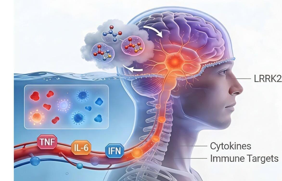
Microglia are a specialized type of immune cell that accounts for about 10% of all cells within the brain and spinal cord. They function by eliminating infectious microbes, dead cells, and aggregated proteins, as well as soluble antigens that may endanger the brain and, during development, also help shape neural circuits enabling specific brain functions.

When microglia don’t function properly, they can trigger neuroinflammation and fail to clear away damaged cells and harmful protein clumps—such as the neurofibrillary tangles and amyloid plaques seen in Alzheimer’s disease. This contributes to numerous neurodegenerative diseases, including Alzheimer’s, Parkinson’s and Huntington’s disease, as well as amyotrophic lateral sclerosis (ALS), multiple sclerosis, and other disorders. In fact, neuroinflammation can occur even before proteins start to form pathogenic aggregates and, in turn, accelerates protein aggregation.

Researchers and drug developers aiming to better understand and target microglia functions in the brain are challenged by the fact that human microglia can only be obtained through biopsies, and rodents’ microglia differ from their human counterparts in many critical features. This supply issue prompted them to work on methods to create microglia in the culture dish using stem cells as a starting point. However, to date, this process has remained inefficient, and requires weeks to complete at significant costs.

Researchers at the Department of Energy’s Lawrence Berkeley National Laboratory (Berkeley Lab) have made a breakthrough in laser technology by using machine learning (ML) to help stabilize a high-power laser.

This advancement, spearheaded by Berkeley Lab’s Accelerator Technology & Applied Physics (ATAP) and Engineering Divisions, promises to accelerate progress in physics, medicine, and energy. The researchers report their work in the journal High Power Laser Science and Engineering.

Scientists know the stomach talks to the brain, but two new studies from Rutgers Health researchers suggest the conversation is really a tug-of-war, with one side urging another bite, the other signaling “enough.”

Together, the papers in Nature Metabolism and Nature Communications trace the first complementary wiring diagram of hunger and satiety in ways that could refine today’s blockbuster weight-loss drugs and blunt their side effects.

One study, led by Zhiping Pang of Robert Wood Johnson Medical School’s Center for NeuroMetabolism, pinpointed a slender bundle of neurons that runs from the hypothalamus to the brainstem.

Breast cancer is the most prevalent malignancy among women worldwide. Phototheranostics—an approach that uses light both to detect and treat cancerous lesions—has drawn growing attention due to its potential advantages, including light-triggered, non-invasive real-time diagnosis and simultaneous in situ therapy.

One promising strategy in light-based cancer treatment is (PTT), which employs photothermal agents—ideally with tumor-targeting capability—to convert light irradiation into localized heat. However, challenges remain in the clinical translation of PTT, particularly the risks of overheating and damaging , as well as the potential failure to effectively ablate tumors.

In a study published in PNAS, a team led by Zhang Pengfei from the Shenzhen Institutes of Advanced Technology (SIAT) of the Chinese Academy of Sciences, in collaboration with Jong Seung Kim from Korea University, Jonathan L. Sessler from the University of Texas at Austin, and Zhou Hui from the Nanjing University of Posts and Telecommunications, developed a dual-laser PTT (DLPTT) strategy for therapy.

Serotonin signaling and gut-immune crosstalk: the microbiome’s role in antitumor immunity.

“…Serotonin transporter inhibits cytotoxic CD8-positive T lymphocyte antitumor immunity by depleting serotonin within the tumor microenvironment…”

“…Serotonin transporter-blocking selective serotonin reuptake inhibitor antidepressants enhance cytotoxic CD8-positive T lymphocyte antitumor immunity and act synergistically with programmed cell death protein 1 immune checkpoint blockade therapy…”

To this end, here…

“…Tumor-infiltrating cytotoxic CD8-positive T lymphocytes were identified as the primary producers and mediators of a local, immunomodulatory serotonin signaling pathway independent of the gastrointestinal tract…”

“…Upon recognition of tumor antigens, tumor-infiltrating cytotoxic CD8-positive T lymphocytes upregulate tryptophan hydroxylase 1, which synthesizes serotonin followed by its release into the tumor microenvironment to enhance T lymphocyte activation via serotonin signaling…”

In short…

Using an algorithm they call the Krakencoder, researchers at Weill Cornell Medicine are a step closer to unraveling how the brain’s wiring supports the way we think and act. The study, published June 5 in Nature Methods, used imaging data from the Human Connectome Project to align neural activity with its underlying circuitry.

Mapping how the brain’s anatomical connections and activity patterns relate to behavior is crucial not only for understanding how the brain works generally but also for identifying biomarkers of disease, predicting outcomes in neurological disorders and designing personalized interventions.

The brain consists of a complex network of interconnected neurons whose collective activity drives our behavior. The structural connectome represents the physical wiring of the brain, the map of how different regions are anatomically connected.