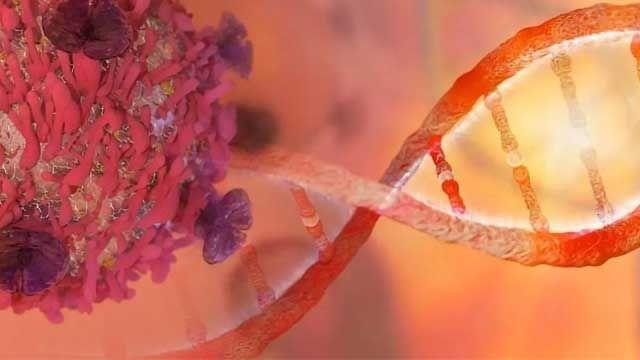
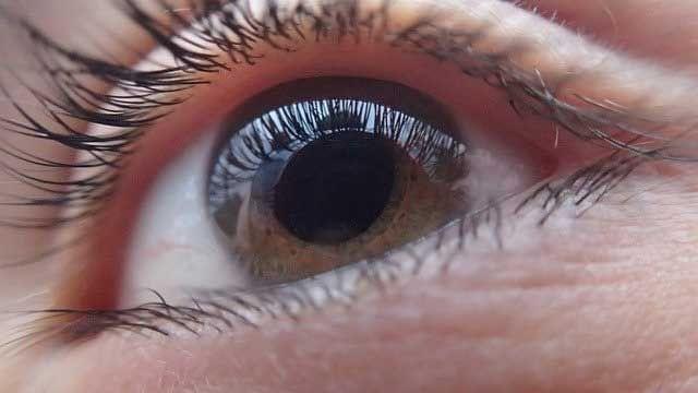
Toggle light / dark theme

Bioengineers and life scientists incorporate hybridoma technology to produce large numbers of identical antibodies, and develop new antibody therapeutics and diagnostics. Recent preclinical and clinical studies on the technology highlight the importance of antibody isotypes for therapeutic efficacy. In a new study, a research team in Netherlands have developed a versatile Clustered Regularly Interspaced Short Palindrome Repeats (CRISPR) and homology directed repair (HDR) platform to rapidly engineer immunoglobin domains and form recombinant hybridomas that secrete designer antibodies of a preferred format, species or isotype. In the study, Johan M. S. van der Schoot and colleagues at the interdisciplinary departments of immunology, proteomics, immunohematology, translational immunology and medical oncology, used the platform to form recombinant hybridomas, chimeras and mutants. The stable antibody products retained their antigen specificity. The research team believes the versatile platform will facilitate mass-scale antibody engineering for the scientific community to empower preclinical antibody research. The work is now published on Science Advances.

Monoclonal antibodies (mAb) have revolutionized the medical field with applications to treat diseases that were once deemed incurable. Hybridoma technology is widely used since 1975 for mAb discovery, screening and production, as immortal cell lines that can produce large quantities of mAbs for new antibody-based therapies. Scientists had generated, validated and facilitated a large number of hybridomas in the past decade for preclinical research, where the mAb format and isotypes were important to understand their performance in preclinical models. Genetically engineered mAbs are typically produced with recombinant technology, where the variable domains should be sequenced, cloned into plasmids and expressed in transient systems. These processes are time-consuming, challenging and expensive, leading to outsourced work at contract research companies, which hamper the process of academic early-stage antibody development and preclinical research.

In its mechanism of action, the constant antibody domains forming the fragment crystallizable – (Fc) domain are central to the therapeutic efficacy of mAbs since they engage with specific Fc receptors (FcRs). Preceding research work had highlighted the central role of Fc in antibody-based therapeutics to emphasize this role. Since its advent, CRISPR and associated protein Cas-9 (CRISPR-Cas9)-targeted genome editing technology has opened multitudes of exciting opportunities for gene therapy, immunotherapy and bioengineering. Researchers had used CRISPR-Cas9 to modulate mAb expression in hybridomas, generate a hybridoma platform and engineer hybridomas to introduce antibody modification. However, a platform for versatile and effective Fc substitution from foreign species within hybridomas with constant domains remains to be genetically engineered.

Imperial College London biomedical materials scientist Molly Stevens teamed up with Massachusetts Institute of Technology biomedical engineer Sangeeta Bhatia to develop the approach, which they think has the potential to help patients in low-resource and rural areas, where available medical technology may be limited. Stevens specializes in low-cost catalyst-based diagnostics and Bhatia works on creating nanosensors that respond to enzymatic activity. The two combined their expertise to create nanoparticle-protein complexes that, once injected, can reveal the presence of disease-related enzymes through a simple urine test.


Sensor turns urine blue in the presence of tumor-related enzymes.

by.

While X-rays can produce harmful radiation, a new technique using laser-induced sound waves provides highly detailed images of the structures in our bodies.
» Subscribe to Seeker!http://bit.ly/subscribeseeker
» Watch more Elements! http://bit.ly/ElementsPlaylist

Photoacoustic imaging is an emerging imaging technique that shoots micro-pulses of laser light at a specimen or body part, which selectively heats up parts of the tissue causing them to expand, and generate waves of pressure – a.k.a. sound waves.

Ultrasonic sensors are situated to capture these microscopic changes, and a processing software then reconstructs the image based on what the sensors “hear.” The speed of the laser can be adjusted depending on what type of tissue one would like to visualize.

The photoacoustic imaging technique is beginning to take off in both the medical and scientific worlds, as it provides us with super clear, incredibly detailed images of the human body and the structures inside it.

Researchers are blurring the distinction between brain and machine, designing nanoelectronics that look, interact, and feel like real neurons. Camouflaged in the brain, this neurotechnology could offer a better way to treat neurodenerative diseases or control prosthetics, interface with computers or even enhance cognitive abilities.

Electrodes implanted in the brain help alleviate symptoms like the intrusive tremors associated with Parkinson’s disease but current probes face limitations due to their size and inflexibility. In a recent paper titled “Precision Electronic Medicine,” published in Nature Biotechnology, Shaun Patel, a faculty member at the Harvard Medical School and Massachusetts General Hospital, and Charles M. Lieber, the Joshua and Beth Friedman University Professor, argue that neurotechnology is on the cusp of a major renaissance. Throughout history, scientists have blurred discipline lines to tackle problems larger than their individual fields.

“The next frontier is really the merging of human cognition with machines,” says Patel. “Everything manifests in the brain fundamentally. All your thoughts, your perceptions, any type of disease.” He and Lieber see mesh electronics as the foundation for these machines, a way to design personalized electronic treatment for just about anything related to the brain. “Today, research focused at the interface between the nervous system and electronics is not only leading to advances in fundamental neuroscience, but also unlocking the potential of implants capable of cellular-level therapeutic targeting,” write the authors in their paper.

AMAZING STUFF, 3D printing is revolutionizing medical and technological science… Respect AEWR wherein we have found the causes and a cure for the pandemic plague mankind has called natural aging when it is the reverse the most unnatural thing on earth to do is age and die. Proven long ago by Science sitting waiting for us to pick it up in the established data of mankind’s humanities… We search for partners-investors to now join us in agiongs end… r.p.berry


The Chicago-based biotech company BIOLIFE4D announced today that it has successfully 3D-bioprinted a mini human heart. The tiny heart has the same structure as a full-sized heart, and the company says it’s an important milestone in the push to create an artificial heart viable for transplant.

Dr. Kevin Strange is the CEO and co-founder of Novo Biosciences, a biotechnology company focused on regenerating the heart and other organs. We recently had the opportunity to interview him about MSI-1436 (trodusquemine), a compound that promotes regeneration in multiple animal models.

What, if anything, happens to existing scar tissue in the presence of MSI-1436?

More detailed studies need to be conducted to fully understand how MSI-1436 impacts existing scar tissue. However, our published work is very encouraging. We induced ischemic injury in the adult mouse heart by permanently ligating the left anterior descending coronary artery. This is a standard heart attack model. Twenty-four hours after ligation of the artery, we began treating with MSI-1436 or vehicle (placebo). Hearts were isolated from mice for histological analysis 3 days and 28 days after injury and collagen deposition (i.e., scarring) was quantified. In hearts isolated after 3 days, the scarring index measured as area was the same, ~40%, in both MSI-1436- and vehicle-treated mice. In other words, there was no difference in the extent of initial scarring in the two groups of animals.