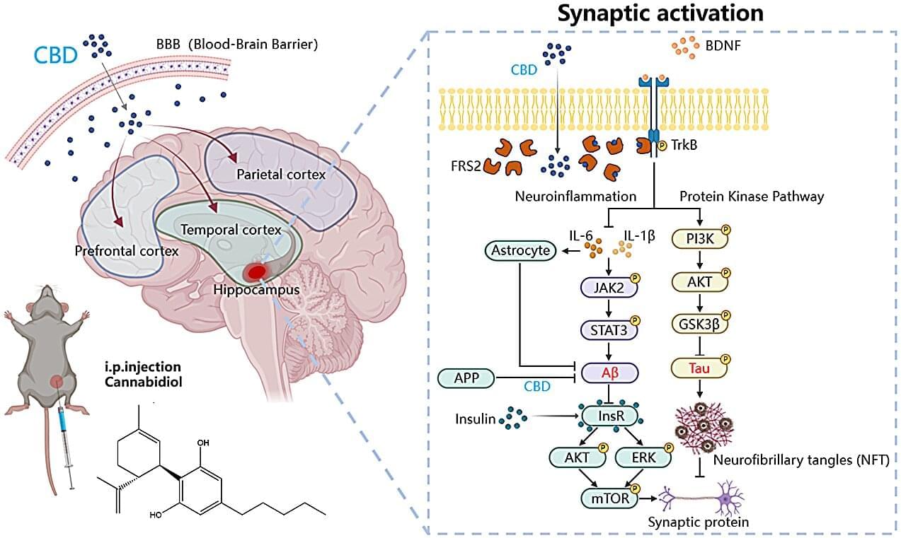
Toggle light / dark theme

Stryker attack wiped tens of thousands of devices, no malware needed

Last week’s cyberattack on medical technology giant Stryker was limited to its internal Microsoft environment and remotely wiped tens of thousands of employee devices.

The organization says in an update on Sunday that all its medical devices are safe to use but electronic ordering systems remain offline, and customers must place orders manually through sales representatives.

Stryker emphasizes that the incident was not a ransomware attack and that the threat actor did not deploy any malware on its systems.

Plasma proteomic signature of frailty in 50,506 adults

Online now: Jia et al. delineate the most comprehensive plasma proteomic landscape of frailty to date and develop proteomic frailty scores that predict multiple diseases and respond to modifiable risk factors. They identify a biphasic pattern of frailty-related proteomic alterations across the lifespan, revealing critical windows that may inform targeted intervention programs.

Compound amino acid synergizes ceftazidime-avibactam to eradicate extracellular and facultative intracellular MDR pathogens

This study demonstrates that the FDA-approved drug 18AA potently resensitizes multidrug-resistant pathogens to ceftazidime-avibactam. It achieves this by activating two bacterial pathways, the inosine-CusS/R-CusC axis and the proton motive force, to promote antibiotic influx, offering a readily translatable strategy against formidable infections.

RNA barcodes fast-track brain connection mapping

“When engineering a computer, you need to know the circuitry of the central processing unit. If you don’t know how everything is wired together, you can’t understand its function, optimize it or fix it when something breaks. We are approaching the brain the same way,” said study leader Boxuan Zhao, a professor of cell and developmental biology at the University of Illinois Urbana-Champaign.

“Our technology enables simultaneous mapping of thousands of neural connections with single-synapse resolution —a capability that doesn’t exist in any current technology. It is directly applicable to understanding circuit dysfunction in neurodegenerative diseases and could provide a platform for developing circuit-guided therapeutic interventions,” he said.

Abstract: Patients with recurrent kidney stone disease stand to benefit from personalized diagnostics

In this Research Article, Ruxandra Bachmann-Gagescu & team integrate blood and urine biochemistry with genetics to improve interpretation of genetic findings in adults with kidney stone disease—the approach has prognostic value, enabling personalized risk assessment.


3Department of Molecular Life Sciences, University of Zurich, Zurich, Switzerland.

4National Center of Competence in Research (NCCR) Kidney. CH, Bern, Switzerland.

5Department of Nephrology and Hypertension, Inselspital, Bern University Hospital, University of Bern, Bern, Switzerland.

ADJUVANT Randomized Controlled Trial: Rationale and Design

ADJUVANT: Testing if IV tirofiban is non-inferior to thrombolysis before endovascular thrombectomy for acute ischemic stroke. Read about the upcoming trial.


BackgroundIntravenous thrombolysis followed by endovascular thrombectomy (EVT) is a first‐line recommended strategy for thrombolysis‐eligible patients with stroke due to acute large‐vessel occlusion. Tirofiban, one of the most used glycoprotein IIb/IIIa receptor inhibitors, has been increasingly advocated to counteract different stages of thrombosis mediated by activated platelets. It is unclear whether tirofiban could be used as an alternative to thrombolysis as adjuvant medicine for EVT. This trial aims to assess whether intravenous tirofiban plus EVT is noninferior to intravenous thrombolysis bridging with EVT in patients with acute ischemic stroke due to large‐vessel occlusion who are eligible for thrombolysis.

Neoantigens and their potential applications in tumor immunotherapy

The incidence of malignant tumors is increasing, the majority of which are associated with high morbidity and mortality rates worldwide. The traditional treatment method for malignant tumors is surgery, coupled with radiotherapy or chemotherapy. However, these therapeutic strategies are frequently accompanied with adverse side effects. Over recent decades, tumor immunotherapy shown promise in demonstrating notable efficacy for the treatment of cancer. With the development of sequencing technology and bioinformatics algorithms, neoantigens have become compelling targets for cancer immunotherapy due to high levels of immunogenicity. In addition, neoantigen-based vaccines have demonstrated potential for cancer therapy, primarily by augmenting T-cell responses. Neoantigens have also been shown to be effective in immune checkpoint blockade therapy.

Plastic bottles transformed into Parkinson’s drug using bacteria

A drug to treat Parkinson’s disease can be made from waste plastic bottles using a pioneering method, a study shows. The approach harnesses the power of bacteria to transform post-consumer plastic into L-DOPA, a frontline medication for the neurological disorder. It is the first time a natural, biological process has been engineered to turn plastic waste into a therapeutic for a neurological disease, researchers say.

Scientists at the University of Edinburgh engineered E. coli bacteria to turn a type of plastic used widely in food and drink packaging—polyethylene terephthalate, or PET—into L-DOPA. The process involves first breaking down PET waste—some 50 million tonnes of which are produced annually—into chemical building blocks of terephthalic acid. Molecules of terephthalic acid are then transformed into L-DOPA by the engineered bacteria through a series of biological reactions.

Using the new technique to produce L-DOPA is more sustainable than traditional methods of making pharmaceuticals, which rely on the use of finite fossil fuels, the team says.

Ultrasound-based technology to deliver large therapeutics into cancer cells

In the study, the authors equipped these microbubbles with synthetic nucleic acid strands designed to bind with specific biochemical receptors that appear on the cell membranes of cancer cells but not healthy cells. They then tried several combinations of ultrasound frequencies and intensities to find the perfect pairing for opening pores in the cell membranes to allow the PROTACs to enter.

Once the optimal settings were identified, the researchers validated the platform by attaching fluorescent molecules to the PROTACs. They conducted separate experiments on cancer cells and healthy cells to compare the delivery efficiency. After a minute of ultrasound exposure, the cells treated with SonoPIN glowed seven times brighter than those treated with traditional PROTAC delivery methods, indicating that they were taking in many PROTACs. This resulted in half of the cancer cells self-destructing, while 99% of the healthy cells remained viable.

Moving forward, the researchers plan to test this approach in mouse models and have already applied for a patent covering the work. By injecting the PROTACs and cancer-seeking microbubbles into their veins and focusing the ultrasound waves on tumor locations, they believe SonoPIN could form a highly potent cancer-killing technology with few side effects. sciencenewshighlights ScienceMission.


Engineers have demonstrated a technique that uses microbubbles and ultrasound to help relatively large cancer drugs enter tumor cells and cause them to self-destruct.

Dubbed “Sonoporation-assisted Precise Intracellular Nanodelivery”—or SonoPIN for short—the technology caused 50% of targeted cancer cells in a benchtop experiment to self-destruct, while leaving 99% of non-targeted cells healthy. The results show promise for precisely delivering a wide variety of large-molecule therapeutics to cells with few off-target effects.

The research appears in the journal Proceedings of the National Academy of Sciences.

Frontiers: Cardiovascular diseases are the leading cause of death in the world

This is partly due to the low regenerative capacity of adult hearts. mRNA therapy is a promising approach under development for cardiac diseases. In mRNA therapy, expression of the target protein is modulated by delivering synthetic mRNA therapy benefits cardiac regeneration by increasing cardiomyocyte proliferation, reducing fibrosis, and promoting angiogenesis. Because mRNA is translated in the cytoplasm, the delivery efficiency of mRNA into the cytoplasm and nucleus significantly affects its therapeutic efficacy. To improve delivery efficiency, non-viral vehicles such as lipid nanoparticles have been developed. Non-viral vehicles can protect mRNA from enzymatic degradation and facilitate the cellular internalization of mRNA. In addition to non-viral vehicles, viral vectors have been designed to deliver mRNA templates into cardiac cells. This article reviews lipid nanoparticles, polymer nanoparticles, and viral vectors that have been utilized to deliver mRNA into the heart. Because of the growing interest in lipid nanoparticles, recent advances in lipid nanoparticles designed for cardiac mRNA delivery are discussed. Besides, potential targets of mRNA therapy for myocardial infarction are discussed. Gene therapies that have been investigated in patients with cardiac diseases are analyzed. Reviewing mRNA therapy from a clinically relevant perspective can reveal needs for future investigations.

Cardiovascular diseases are a group of diseases related to heart muscles, blood vessels, and valves. The death caused by cardiovascular diseases worldwide in 2019 was 17.9 million, which accounts for approximately 30% of total death in the year (DofE and SAPD, 2019; World Heath Organization, 2021). Myocardial infarction and strokes result in over 80% of deaths from cardiovascular diseases. Percutaneous coronary intervention treatment has significantly lowered mortality after acute myocardial infarction. However, the cardiac function will be permanently impaired. Newborn mammals can regenerate the injured heart, but this regenerative capacity disappears in adults (Porrello et al., 2011; Ye et al., 2018). The declined regenerative capacity in aged hearts is partly due to decreased cardiomyocyte proliferation, lowered angiogenesis, and increased fibrosis (Rivard et al., 1999; Senyo et al., 2012; Notari et al., 2018).

/* */