For decades, people have arranged to freeze their bodies after death, dreaming of resurrection by advanced future medicine.


In the largest observational study to date on “SuperAgers” — people in their 80s who have brains as sharp as those 30 years younger — researchers in Spain found key differences in lifestyle that may contribute to these older adults’ razor-sharp minds.
SuperAgers in the study had more gray matter in parts of the brain related to movement, and they scored higher on agility, balance and mobility tests than typical older adults — even though the physical activity levels of the two groups were the same.
“Though superagers report similar activity levels to typical older people, it’s possible they do more physically demanding activities like gardening or stair climbing,” said senior author Bryan Strange, director of the Laboratory for Clinical Neuroscience at the Technical University of Madrid, in a statement.

Immunotherapy could be the “shot in the arm” you need to treat your allergies.
ARLINGTON HEIGHTS, Ill. (August 7, 2019) – You may think you can’t tell the difference between the symptoms caused by spring, summer and fall allergies. They all usually involve sneezing, sniffling, itchy eyes and a runny nose. And while symptoms for each Allergies are inappropriate or exaggerated reactions of the immune system to substances that, in the majority of people, cause no symptoms. Symptoms of the allergic diseases may be caused by exposure of the skin to a chemical, of the respiratory system to particles of dust or pollen (or other substances), or of the stomach and intestines to a particular food. rel= tooltip allergy season may be similar, the treatment can look very different, particularly if Immunotherapy is a form of preventive and anti-inflammatory treatment of allergy to substances such as pollens, house dust mites, fungi, and stinging insect venom.

So why, almost 30 years on from Dolly, haven’t humans been cloned yet? Is it primarily for ethical reasons, are there technological barriers, or is it simply not worth doing?
Related: What are the alternatives to animal testing?
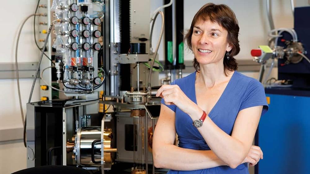
When Nicola Spaldin began studying natural sciences at the University of Cambridge in 1988, she planned on becoming a physicist, but then quickly reconsidered. “After about the second lecture I completely changed my mind,” she recalls. “I thought ‘I’m absolutely not clever enough to be a physicist.’ Everybody was very brilliant and I was not.”
Yet it seems Spaldin was vastly underestimating herself. Now a professor of materials science at ETH Zurich, she won two major awards for physics last year: the EPS Europhysics Prize and the Hamburg Prize for Theoretical Physics. Both accolades cited Spaldin’s pioneering work on the theory of magnetoelectric multiferroics – materials that are both ferromagnetic and ferroelectric. These properties are rarely found together, making it very difficult to engineer substances with both, but they have many exciting potential applications, from microelectronics to medicine.
\r \r
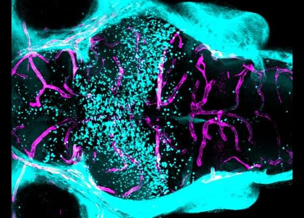
A new study in mice by scientists at St. Jude Children’s Research Hospital reveals that immune and tumor cells compete over glutamine. The researchers also identified a molecular pathway that could serve as a potential drug target to achieve the same effect.
The findings are published in Nature in an article titled, “SLC38A2 and glutamine signaling in cDC1s dictate anti-tumor immunity.”
“Cancer cells evade T cell-mediated killing through tumor–immune interactions whose mechanisms are not well understood,” the scientists wrote. “Dendritic cells (DCs), especially type-1 conventional DCs (cDC1s), mediate T cell priming and therapeutic efficacy against tumors. DC functions are orchestrated by pattern recognition receptors, although other signals involved remain incompletely defined. Nutrients are emerging mediators of adaptive immunity, but whether nutrients affect DC function or communication between innate and adaptive immune cells is largely unresolved. Here we establish glutamine as an intercellular metabolic checkpoint that dictates tumor–cDC1 crosstalk and licenses cDC1 function in activating cytotoxic T cells.”

China: A recent article published in Clinical, Cosmetic and Investigational Dermatology reports the case of mycobacterium marinum skin infection in the left upper limb of a female patient with chronic idiopathic myelofibrosis during ruxolitinib treatment.
“Our case illustrates the diversity of skin infections that may occur during JAK (Janus kinase) inhibitors treatment, and the need for clinical attention to atypical mycobacterial skin infections cannot be ignored,” Xiaonan Chen, Affiliated Hospital of Weifang Medical University, Weifang, People’s Republic of China, and colleagues wrote in their case study.
Mycobacterium marinum is an atypical bacterium, and skin infections that result from it are relatively rare, usually occurring in homemakers who clean and prepare fish for food and workers engaged in seafood processing. The infection often occurs after fish spines, scales, etc., puncture the skin. The JAK/STAT signalling pathway is closely linked with the human immune response to infections. Therefore, JAK inhibitors may induce and exacerbate various conditions in clinical practice.


Simply the smell of seafood can make those with an allergy to it violently ill—and therefore more likely to avoid it. The same avoidance behavior is exhibited by people who develop food poisoning after eating a certain meal.
Scientists have long known that the immune system played a key role in our reactions to allergens and pathogens in the environment, but it was unclear whether it played any role in prompting these types of behaviors towards allergic triggers.
According to Yale-led research published July 12 in the journal Nature, it turns out that the immune system plays a crucial role in changing our behaviors.