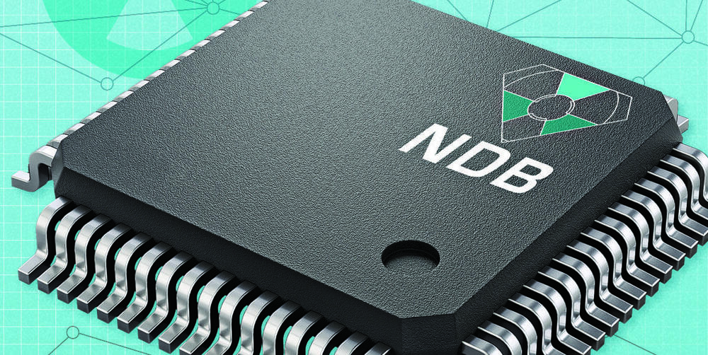
In less than two years, you might be able to buy a smartwatch —powered with a radioactive diamond battery—that will outlive you and your progeny for generations.
The potentially game-changing battery comes from the San Francisco–based startup Nano Diamond Battery (NDB), which lauds its namesake “high-power diamond-based alpha, beta, and neutron voltaic battery” for its ability to give devices “life-long and green energy.” Imagine: Just one battery could power your insulin pump or pacemaker for your entire life (with loads of time to spare). Or it could provide the juice for a space rover, collecting Mars regolith samples for decades without any human assistance.
Those are ambitious goals. So, could NDB’s bold claims actually become reality?
Culinary Herbs & Spices For Health, Wellness & Longevity — Dr. Hamed Faridi Ph.D., Executive Director, McCormick Science Institute
Dr. Hamed Faridi, Ph.D. is the founder of Faridi Strategy Group LLC and serves as the Executive Director of the McCormick Science Institute (https://www.mccormickscienceinstitute.com/).
Hamed is renowned as an innovative food industry leader, business executive, strategist, and board director. He is a visionary leader who conceives and implements innovative approaches — often using technology — to create and sustain business growth in the highly competitive food manufacturing industry. Hamed is known as someone who creates “momentum” and superior customer intimacy.
Hamed is a sought-after consultant and frequent industry speaker with valuable perspectives on the food industry and the “future of food”. He has a reputation for developing strong and trusting relationships with CEOs, executive leaders, industry peers, and board directors. He is considered an effective communicator, a good listener, and a team-mate whose insights are valued. He has significant experience in the technology, health care, and food / flavor industries.
Hamed has served on boards of directors of several organizations including Maryland University of Integrative Health, St. Joseph Medical Center, and the International Association of Cereal Chemists. He has been a director and president of both the Flavor & Extract Manufacturers Association and the American Association of Cereal Chemists. He has served on the partnership committee of a McCormick joint venture and on the advisory boards of the food science departments of four different universities.
About the Bioprint FirstAid Handheld Bioprinter capabilities.
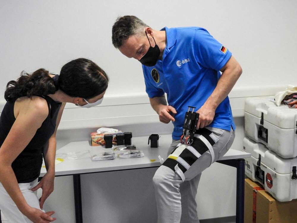
Astronauts on the International Space Station (ISS) are testing 3D bioprinted bandages made of their own cells that could be used to better heal flesh wounds in space.
The German Space Agency (DLR) is leading the experiment which was launched to the ISS at the end of December 2021 on SpaceX’s 24th commercial resupply mission. The payload contained the BioPrint FirstAid Handheld Bioprinter, which is designed to hold cells from astronauts within a bioink that can be used to apply bandages to wounds when needed.
While the experiment offers a promising tool for wound healing in space environments, it could also provide significant benefits back on earth, too.
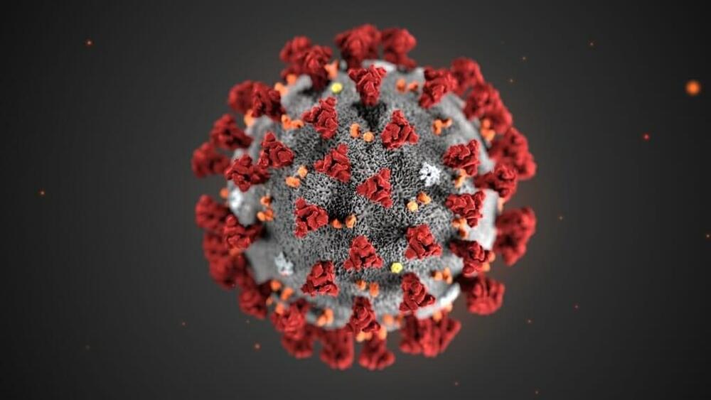
Viruses can be wily adapters, changing their identities to find new hosts and thwart efforts to stop them. That’s why University of Wisconsin-Madison researchers and their collaborators are making progress toward developing universal vaccines against some the planet’s most harmful pathogens, including the virus family responsible for the COVID-19 pandemic.
Last fall, the National Institutes of Health announced it was investing in three teams working to develop a vaccine that would simultaneously work against a broad range of coronaviruses. Among them is a research collaboration, the Pan-Coronavirus Vaccine consortium, led by UW-Madison School of Veterinary Medicine Professor of Pathobiological Sciences Yoshihiro Kawaoka.
“This pan-coronavirus vaccine is basically preparing for the future,” Kawaoka says.
If the world already had a pan-coronavirus vaccine in March 2020, it could have served as a mitigation tool until vaccines specific to SARS-CoV-2 could be developed.
Athletes headed to the Beijing Olympic Winter Games are making final travel preparations, including keeping in line with China’s health measures on the “My 2022″ smartphone app. However, inadequate encryption measures within the app can leave Olympians, journalists and sports officials vulnerable to hackers, privacy breaches, and surveillance, according to a cybersecurity report by the Citizen Lab obtained exclusively by DW. Additionally, the IT forensic specialists found that the app includes a censorship keyword list. The findings come as international concern over digital safety at the Games mounts. Germany, Australia, UK and US have urged their athletes and National Olympic Committees to leave their personal phones and laptops behind and to travel with special devices over fears of digital espionage. The Dutch Olympic Committee outright banned its athletes from bringing personal phones and laptops due to surveillance concerns.
In the Olympic Playbook for athletes and team officials, the International Olympic Committee states that the “My 2022″ app is “in accordance with international standards and Chinese law.” But based on its findings, Citizen Lab concludes that the insecure transmission of personal information “may constitute a direct violation of China’s privacy laws.” This is because China’s data protection laws require that a person’s health and medical records held digitally be transmitted and stored in an encrypted manner. Citizen Lab’s findings also raise questions concerning two Western tech giants that carry the “My 2022″ app: Apple and Google. “Both Apple’s and Google’s policies forbid apps to transmit sensitive data without proper encryption, so Apple and Google will need to determine whether the app’s unresolved vulnerabilities warrant delisting,” Citizen Lab’s Knockel told DW. The Beijing Organizing Committee has stood by its app, however, saying it “passed the examination” of international mobile application markets such as Google, Apple and Samsung.“We have taken measures such as personal information encryption in the app to ensure privacy security,” the committee said Monday to Xinhua News Agency.
The Winter Games, which kicks off on February 4, marks the second Olympic Games during the COVID-19 pandemic. Just as at the Tokyo Summer Games, tracking athletes’ health is required. According to the official Playbook of the International Olympic Committee (IOC), athletes, coaches, reporters and sports officials, as well as thousands of local staff, are required to put their information into either the “My 2022″ smartphone app or website. The app, which was developed in China, is designed to monitor the health of all attendees and staff as well as trace possible COVID-19 infections. Passport data and flight information must be entered into the app. Sensitive medical information related to possible COVID-19 symptoms are also required, such as whether a person had a fever, fatigue, headaches, a dry cough, diarrhea or a sore throat. Those coming from abroad must start entering health data 14 days before arriving in the country. Many countries use a contact tracing app to help combat the pandemic. But “My 2022″ combines contact tracing with other services: It regulates access to events, acts as a visitor’s guide with information on sporting venues and tourist services, as well as providing chat functions (text and audio), news feeds and file transfers.
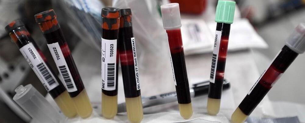
The human body did not evolve to handle life in space, and it shows in our very blood.
Since our species first started to spend extended periods of time beyond our planet, researchers have noticed a curious and consistent loss of red blood cells among astronauts.
The phenomenon is called ‘space anemia’, and until recently, its cause was a mystery. Some experts have argued space anemia is only a short-term phenomenon – a brief compensation for the fluid changes in our bodies under microgravity.
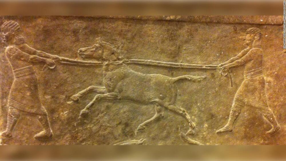
Bronze Age bioengineers created the earliest hybrid animal — a majestic horselike creature known as a kunga that had a donkey mom, a Syrian wild ass for a father and lived 4,500 years ago, according to new research based on the sequencing of DNA from the animal’s skeleton.
Descriptions and imagery in Mesopotamian art and texts portray a powerful animal that pulled war wagons into battle and royal vehicles in parades. Its true identity, however, had long puzzled and divided archaeologists. Domesticated horses didn’t arrive in the region, sometimes referred to as the Fertile Crescent, until 4,000 years ago.
One possible application of interest to researchers is bone healing. The idea is that the soft material, powered by the electroactive polymer, will be able to maneuver in spaces of complicated bone fractures and expand. When the material hardens, it can form the basis for building new bones. In their study, the researchers demonstrate that the material can wrap itself around chicken bones, and the artificial bone that develops later grows along with the animal’s bone. The developed biohybrid variable-stiffness actuators can be used in soft (micro-)robotics and as potential tools for bone repair or bone tissue engineering.
“By controlling how the material turns, we can make the microrobot move in different ways, and also affect how the material unfurls in broken bones. We can embed these movements into the material’s structure, making complex programs for steering these robots unnecessary”, says Edwin Jager.
Tech companies continued to draw criticism for their roles in political and social scandals, most notably when whisteblower and former Facebook employee Frances Haugen testified to lawmakers. Undeterred, Facebook rebranded itself Meta and said it would now focus on building the metaverse. Twitter CEO Jack Dorsey stepped down and likewise changed the name of his company Square to Block in a not-so-subtle nod to the blockchain.
Meanwhile, volatile cryptocurrencies set new records, their prices jumping and crashing on a tweet. NFTs, a once-obscure type of cryptoasset, went on an eye-watering tear as redditors pushed meme stocks skyward. It was also the year of ever-bigger AI. Machine learning models surpassed a trillion parameters, designed computer chips, and tackled practical problems in biology, math, and chemistry. Elsewhere, billionaires went to space, regular folks bought 3D printed houses, fusion power attracted billions in investment, gene editing trials hit their stride, and “flying car” companies hit the New York Stock Exchange.
For this year’s list of fascinating stories in tech and science, we sifted our Saturday posts and selected articles that looked back to where it all began, glanced ahead to what’s coming, or otherwise stood out from the chatter to stand the test of time.