In this episode, I am joined by Dr. David Sinclair, tenured Professor of Genetics at Harvard Medical School and an expert researcher in the field of longevity. Dr. Sinclair is also the author of the book Lifespan: Why We Age & Why We Don’t Have To, and the host of the Lifespan Podcast, which launches January 5, 2022. In this interview, we discuss the cellular and molecular mechanisms of aging and what we all can do to slow or reverse the aging process. We discuss fasting and supplementation with resveratrol, NAD, metformin, and NMN. We also discuss the use of caffeine, exercise, cold exposure, and why excessive iron load is bad for us. We discuss food choices for offsetting aging and promoting autophagy (clearance of dead cells). And we discuss the key blood markers everyone should monitor to determine your biological versus chronological age. We also discuss the future of longevity research and technology. This episode includes lots of basic science and specific, actionable protocols, right down to the details of what to do and when. By the end, you will have in-depth knowledge of the biology of aging and how to offset it. #HubermanLab #DavidSinclair #Longevity
Category: biotech/medical – Page 1,364

AI generates proteins with exceptional binding strengths
A new study in Nature reports an AI-driven advance in biotechnology with implications for drug development, disease detection, and environmental monitoring. Scientists at the Institute for Protein Design at the University of Washington School of Medicine used software to create protein molecules that bind with exceptionally high affinity and specificity to a variety of challenging biomarkers, including human hormones.
Notably, the scientists achieved the highest interaction strength ever reported between a computer-generated biomolecule and its target.
Senior author David Baker, professor of biochemistry at UW Medicine and Howard Hughes Medical Institute investigator, emphasized the potential impact: “The ability to generate novel proteins with such high binding affinity and specificity opens up a world of possibilities, from new disease treatments to advanced diagnostics.”
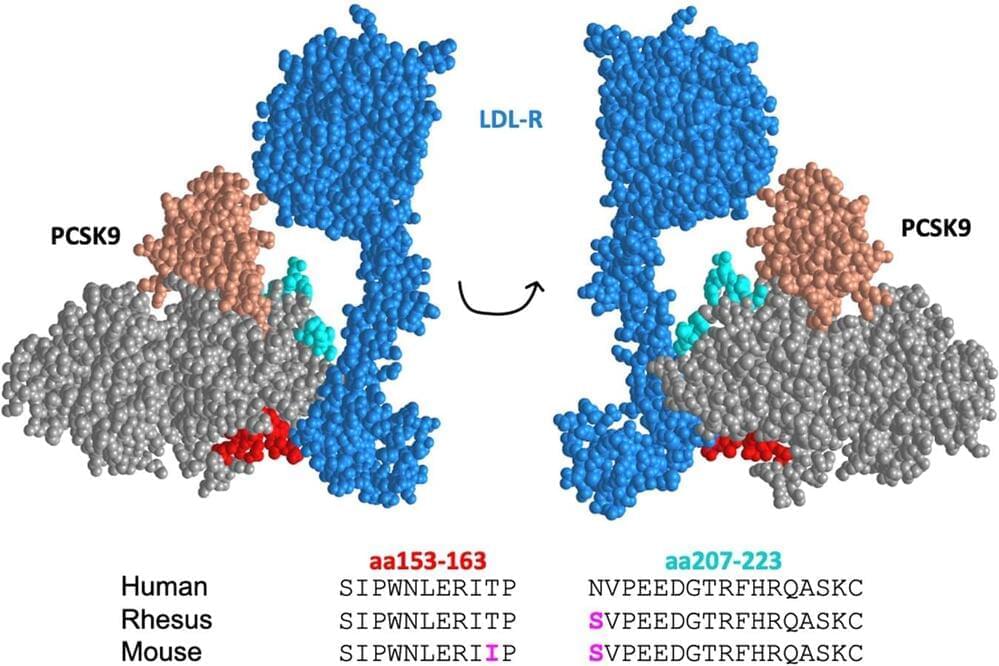
The future of heart health: Researchers develop vaccine to lower cholesterol
Nearly two in five U.S. adults have high cholesterol, according to the Centers for Disease Control and Prevention (CDC). Untreated, high cholesterol can lead to heart disease and stroke, which are two of the top causes of death in the U.S. Worldwide; cardiovascular diseases claim nearly 18 million lives every year, according to the World Health Organization.
A new vaccine developed by researchers at The University of New Mexico School of Medicine could be a game-changer, providing an inexpensive method to lower “bad” LDL cholesterol, which creates dangerous plaques that can block blood vessels.
In a recent study published in npj Vaccines, a team led by Bryce Chackerian, Ph.D., Regents’ Professor in the Department of Molecular Genetics & Microbiology, reported the vaccines lowered LDL cholesterol almost as effectively as an expensive class of drugs known as PCSK9 inhibitors.

Brain Autopsies Suggest a New Culprit Behind Alzheimer’s Disease
Analysis of human brain tissue reveals differences in how immune cells behave in brains with Alzheimer’s disease compared to healthy brains, indicating a potential new treatment target.
University of Washington-led research, published in August, discovered microglia in the brains of people with Alzheimer’s disease were in a pre-inflammatory state more frequently, making them less likely to be protective.
Microglia are immune cells that help keep our brains healthy by clearing waste and preserving normal brain function.
Vertex Pharmaceuticals
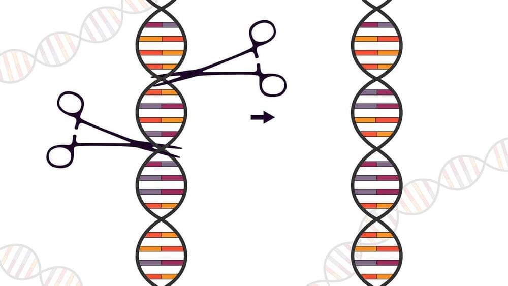
Our research at Vertex is built upon two strong pillars: biology and therapeutic innovation. We continue to fill our drug discovery toolbox with cutting-edge tools and technologies. One of these tools is CRISPR/Cas9 gene editing. Watch this video to learn about how CRISPR/Cas9 gene editing works and how it can be used in therapeutic development.\
For company updates and to learn more about Vertex Pharmaceuticals, follow us on Twitter (/ vertexpharma, YouTube (/ @vertexpharmaceuticalsglobal) and LinkedIn (/ vertex-pharmaceuticals, or visit our website at www.vrtx.com.

Instagram-Famous Plastic Surgeon Sued by Patients: ‘He’s a Danger to the Community’
Several women have filed medical malpractice lawsuits against a southern California plastic surgeon well known on Instagram, alleging that they endured botched surgeries, unexpected scarring and serious infections.
As reported by the Los Angeles Times, the series of lawsuits allege that Dr. Arian Mowlavi degraded the women by requiring them to take off all of their clothes during an examination, touching their bodies without consent and attempting to sell them additional procedures like breast augmentations and body sculpting.
Mowlavi, who was based in Laguna Beach, called himself “Dr. Laguna” and a “renowned body sculptor” while charging 10s of thousands for procedures that his patients allege were done sloppily or, in some cases, not by Mowlavi at all.

Long COVID Rate in Africa Is Almost 50% of Cases, Researchers Warn
Almost half the people known to have contracted COVID-19 in Africa are now living with the effects of long COVID, according to a comprehensive review of previous studies and analysis, covering data from a total of 29,213 people.
Officially, long COVID means persisting symptoms three months after infection with the SARS-CoV-2 virus. Worldwide, at least 10 percent of infected people are thought to suffer from the condition, with recently published research indicating higher rates in multiple countries.
Incidence rates in Africa, though, are well above any of these other estimates. Why that is the case is unclear. In low-income countries, estimates of the incidence of long COVID vary greatly, due to hidden infections and the difficulty of accessing tests.
Improving Gut Health with Microbiome and Probiotics
View show notes here: https://bit.ly/3GJjQKz Become a member to receive exclusive content: https://peterattiamd.com/subscribe/ Sign up to receive Peter’s email newsletter: https://peterattiamd.com/newsletter/ Colleen Cutcliffe is an expert in molecular biology and co-founder of Pendulum Therapeutics, a company working to develop treatments for a variety of diseases by targeting the microbiome. In this episode, Colleen delves into the complexity of the microbiome, how it is tested, and how it changes over time. She explores how probiotics, prebiotics, and postbiotics affect the gut and makes a compelling case that well-developed products have the potential not only to enhance gut health but also to positively influence overall metabolic well-being. Colleen emphasizes the significance of a high-fiber diet in sustaining a thriving gut microbiome, shares insights on minimizing microbiome damage during antibiotic use, provides tips for fostering and preserving a healthy gut, and much more. We discuss: 0:00:00 — Intro 0:00:34 — Colleen’s background and current focus 0:03:08 — The basics of the microbiome 0:12:37 — The study of the human microbiome 0:17:42 — Categories of bacteria, and the implications on health of the rapid evolution of bacteria 0:27:51 — Methods for measuring and understanding the microbiome, and key indicators of microbiome health 0:39:52 — The important role of fiber for promoting gut health through the production of butyrate 0:47:21 — The case for manipulating gut bacteria via fecal microbiota transplant (FMT) 0:53:25 — Dynamics of the microbiome: the gut-brain connection and how antibiotics, nutrition, stress, and more impact the microbiome’s diversity and function 0:59:16 — Factors that influence the vaginal microbiom 1:03:46 — The effect of gut microbes on obesity and challenges with fecal transplants in people 1:06:25 — Beneficial strains of gut bacteria and strains commonly found in probiotics 1:16:35 — The difference between a probiotic and prebiotic, and how CFUs are a measure of the “active ingredient” 1:21:47 — Considerations about how probiotic strains are produced, and more on the meaning of CFU 1:31:12 — Mitigating the effect of antibiotics on the microbiome 1:39:58 — What do we know about the effect of artificial sweeteners on the gut microbiome? 1:47:02 — Why Akkermansia is a keystone strain with implications for metabolic health and an individual’s response to dietary interventions 1:58:14 — The essential steps necessary to develop a robust probiotic for optimal health support 2:01:45 — How Akkermansia helps control blood glucose, and potential implications of Akkermansia in weight loss, diabetes management, and more 2:22:46 — Pendulum Therapeutics’ commitment to rigorous product develop 2:29:54 — Details about the probiotic “Glucose Control” and other probiotics developed by Pendulum Therapeutics 2:38:43 — Further studies of Akkermansia that have been proposed or are underway ——– About: The Peter Attia Drive is a deep-dive podcast focusing on maximizing longevity, and all that goes into that from physical to cognitive to emotional health. With over 70 million episodes downloaded, it features topics including exercise, nutritional biochemistry, cardiovascular disease, Alzheimer’s disease, cancer, mental health, and much more. Peter Attia is the founder of Early Medical, a medical practice that applies the principles of Medicine 3.0 to patients with the goal of lengthening their lifespan and simultaneously improving their healthspan.