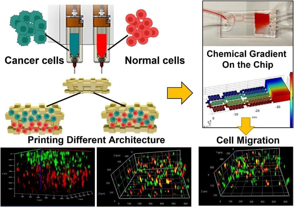
Fluicell, a bioprinting firm based in Sweden, has launched its new high-precision 3D printer, the Biopixlar AER.
Intended as a successor to the original Biopixlar, the device is Fluicell’s second single-cell 3D bioprinting system. The company has designed its latest machine to be as compact and accessible as possible, and claims that it’s the world’s first microfluidic bioprinter that fits inside a standard flow hood or biosafety cabinet. This enables users to easily integrate it with other in vitro and 3D cell culture technologies.
Victoire Viannay, CEO of Fluicell, said, “With Biopixlar AER, we have reached a new important milestone and we can now offer a pioneering product, fully tailored to meet current and future needs in the rapidly accelerating life science and research sector.”
Medcalf: Because you’re moving away from the economics of scale to closer to the clinic, the batches are smaller and some of the traditional paradigms for quality assurance, such as proof of sterility, are harder to arrange. Thus, you need to have a manufacturing system that includes quality assurance within the system itself.
Automation is often presented as a way to remove the single largest source of infective risk, i.e. the human operator. For example, the self-sterilizing reusable units being developed at the University of Osaka under Professor Masahiro Kino-oka allow small-scale production with a high degree of confidence in the aseptic management of the environment.
Another challenge is defining a product that has variable characteristics. The main reason for decentralizing is to allow customization to a patient, which means you need to have a hierarchy of levels of specification. For example, with bioprinting, which also produces a customized product, you need to define bulk properties, but you also need to set constraints around how it’s anchored or implanted into the patient.
Bioprinting is widely applicable to develop tissue engineering scaffolds and form tissue models in the lab. Materials scientists use this method to construct complex 3D structures based on different polymers and hydrogels; however, relatively low resolution and long fabrication times can result in limited procedures for cell-based applications.
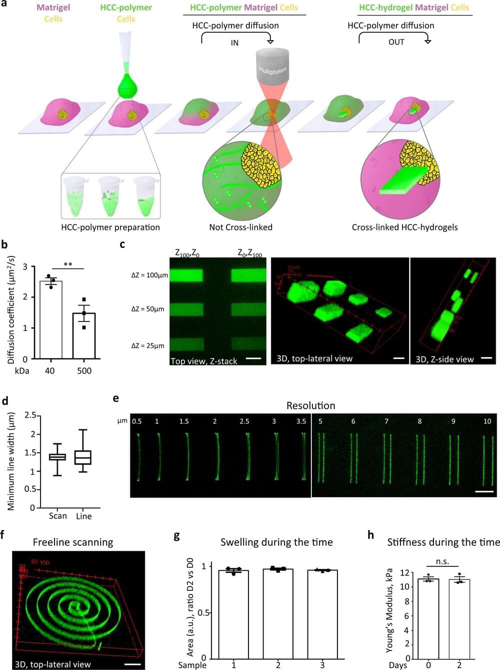
In a new report now available in Nature Asia Materials, Byungjun Lee and a team of scientists in mechanical engineering at Seoul National University, Seoul, Korea, presented a 3D hybrid-micromesh assisted bioprinting method (Hy-MAP) to combine digital light projection, 3D printed micromesh scaffold sutures, together with sequential hydrogel patterning. The new method of bioprinting offered rapid cell co-culture via several methods including injection, dipping and draining. The work can promote the construction of mesoscale complex 3D hydrogel structures across 2D microfluidic channels to 3D channel networks.
Lee et al. established the design rules for Hy-MAP printing via analytical and experimental investigations. The new method can provide an alternative technique to develop mesoscale implantable tissue engineering constructs for organ-on-a-chip applications.
A research team from Utrecht University has successfully fabricated working livers using a newly developed ultrafast volumetric 3D bioprinting method.
By means of visible light tomography, the volumetric bioprinting method enabled the successful printing of miniature stem cell units by making the cells “transparent”, which meant they retained their resolution and ability to perform biological processes.
Printed in less than 20 seconds, the liver units were able to perform key toxin elimination processes mimicking those that natural livers perform in our bodies, and could open new opportunities for regenerative medicine and personalized drug testing.
Named Suspended Layer Additive Manufacturing (SLAM), the approach enables the printing of a novel biomaterial that accurately simulates the structure of mammalian skin.
In fact, according to the researchers, the biomaterial is the first of its kind to simulate all three of the major layers found in skin – the hypodermis, the dermis, and the epidermis – making it a unique tri-layered skin equivalent. Early experiments suggest that the 3D bioprinted skin can be placed at the site of a wound to induce healing, reducing scar tissue in the process.
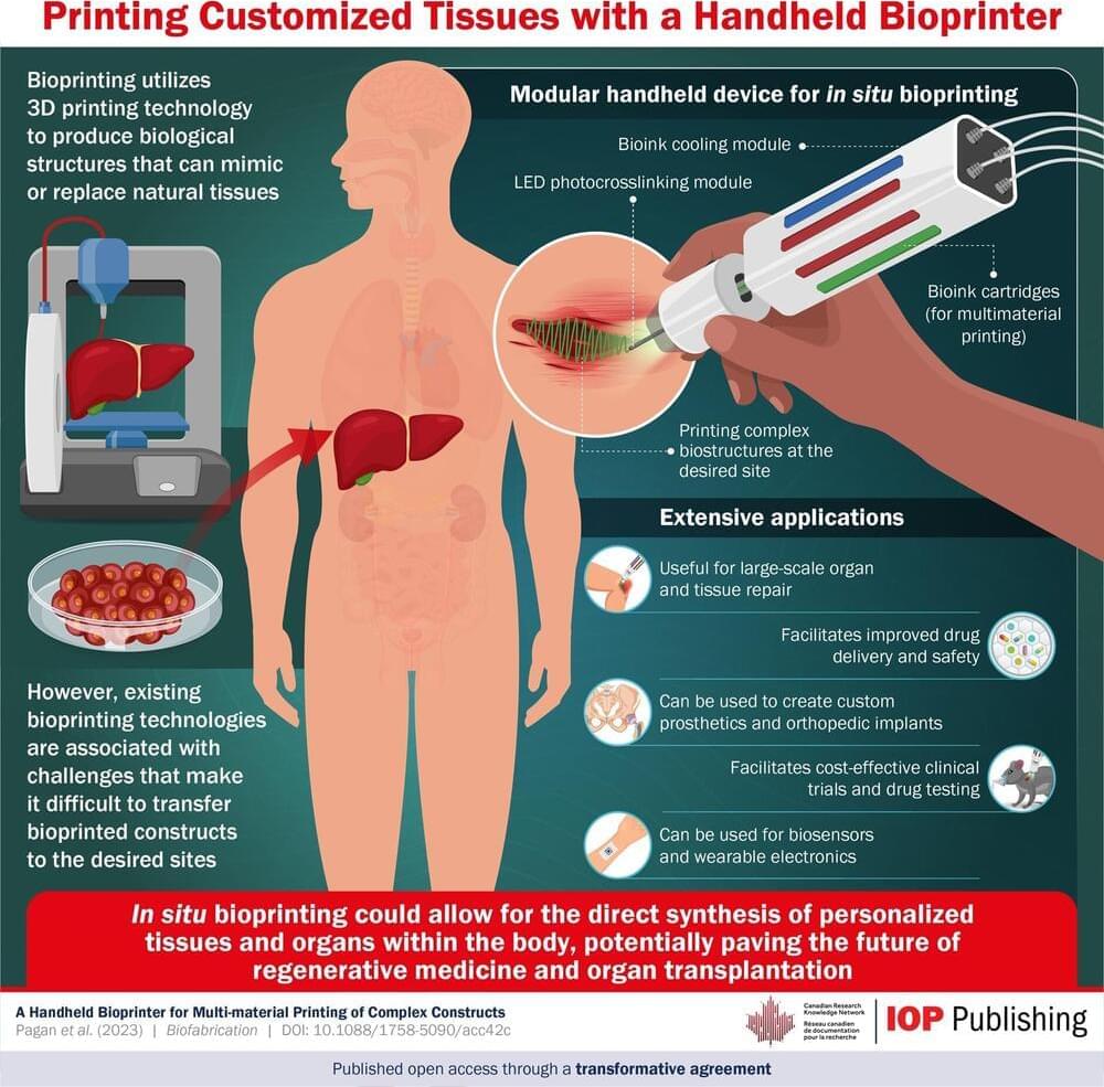
About the Bioprint FirstAid Handheld Bioprinter capabilities.
Astronauts on the International Space Station (ISS) are testing 3D bioprinted bandages made of their own cells that could be used to better heal flesh wounds in space.
The German Space Agency (DLR) is leading the experiment which was launched to the ISS at the end of December 2021 on SpaceX’s 24th commercial resupply mission. The payload contained the BioPrint FirstAid Handheld Bioprinter, which is designed to hold cells from astronauts within a bioink that can be used to apply bandages to wounds when needed.
While the experiment offers a promising tool for wound healing in space environments, it could also provide significant benefits back on earth, too.
Bio-Printing Complex Human Tissues & Organs — Dr. Anthony Atala, MD — Director, Wake Forest Institute for Regenerative Medicine, Wake Forest School of Medicine, Wake Forest University.
Dr. Anthony Atala, MD, (https://school.wakehealth.edu/Faculty/A/Anthony-Atala) is the G. Link Professor and Director of the Wake Forest Institute for Regenerative Medicine, and the W. Boyce Professor and Chair of Urology.
A practicing surgeon and a researcher in the area of regenerative medicine, fifteen applications of technologies developed Dr. Atala’s laboratory have been used clinically. He is Editor of 25 books and 3 journals, has published over 800 journal articles, and has received over 250 national and international patents. Dr. Atala was elected to the Institute of Medicine of the National Academies of Sciences, to the National Academy of Inventors as a Charter Fellow, and to the American Institute for Medical and Biological Engineering.
Dr. Atala is a recipient of the US Congress funded Christopher Columbus Foundation Award, bestowed on a living American who is currently working on a discovery that will significantly affect society; the World Technology Award in Health and Medicine, for achieving significant and lasting progress; the Edison Science/Medical Award for innovation, the R&D Innovator of the Year Award, and the Smithsonian Ingenuity Award for Bioprinting Tissue and Organs. Dr. Atala’s work was listed twice as Time Magazine’s Top 10 medical breakthroughs of the year, and once as one of 5 discoveries that will change the future of organ transplants. He was named by Scientific American as one of the world’s most influential people in biotechnology, by U.S. News & World Report as one of 14 Pioneers of Medical Progress in the 21st Century, by Life Sciences Intellectual Property Review as one of the top key influencers in the life sciences intellectual property arena, and by Nature Biotechnology as one of the top 10 translational researchers in the world.
Dr. Atala has led or served several national professional and government committees, including the National Institutes of Health working group on Cells and Developmental Biology, the National Institutes of Health Bioengineering Consortium, and the National Cancer Institute’s Advisory Board. He is a founding member of the Tissue Engineering Society, Regenerative Medicine Foundation, Regenerative Medicine Manufacturing Innovation Consortium, Regenerative Medicine Development Organization, and Regenerative Medicine Manufacturing Society.
A major obstacle to widespread study and clinical use of 3D tissues is their short shelf-life, which may be anywhere from a just few hours to a few days. As in the case of an organ transplant, a bioprinted tissue must be transported rapidly to the location where it is needed, or it will not be viable. In the journal Matter on December 21st, researchers at Brigham and Women’s Hospital and Harvard Medical School describe their work combining 3D bioprinting with cryopreservative techniques to create tissues which can be preserved in a freezer at-196°C and thawed within minutes for immediate use.
“For conventional bioprinting, there is basically no shelf life. It’s really just print, and then use, in most cases,” says lead author Y. Shrike Zhang (@shrikezhang), a biomedical engineer at Brigham and Women’s Hospital. “With cryobioprinting, you can print and store in the frozen state for basically as long as you want.”
The use of 3D bioprinting to create artificial human tissue first appeared twenty years ago. As in conventional 3D printing, an ink is extruded layer by layer through a nozzle into a pre-specified shape. In the case of bioprinting, the ink is typically made up of a gelatin-like scaffolding embedded with living cells. Cryobioprinting works the same way, except the printing is performed directly onto a cold plate held at temperatures down to-20°C. After the tissues are printed, they are immediately moved to cryogenic conditions for long-term storage.
The 24thSpaceX cargo resupply services mission, targeted to launch in late December from NASA’s Kennedy Space Center in Florida, carries scientific research and technology demonstrations to the International Space Station. The experiments aboard include studies of bioprinting, crystallization of monoclonal antibodies, changes in immune function, plant gene expression changes, laundering clothes in space, processing alloys, and student citizen science projects.