Toggle light / dark theme

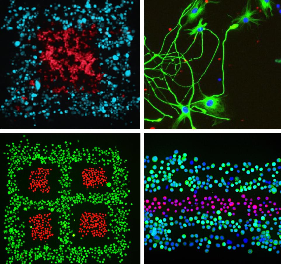
A new study from North Carolina State University shows a reproducible way of studying cellular communication among varied types of plant cells by “bioprinting” these cells via a 3D printer. Learning more about how plant cells communicate with each other—and with their environment—is key to understanding more about plant cell functions and could ultimately lead to creating better crop varieties and optimal growing environments.

The researchers bioprinted cells from the model plant Arabidopsis thaliana and from soybeans to study not just whether plant cells would live after being bioprinted—and for how long—but also to examine how they acquire and change their identity and function.

“A plant root has a lot of different cell types with specialized functions,” said Lisa Van den Broeck, an NC State postdoctoral researcher who is the first author of a paper describing the work. “There are also different sets of genes being expressed; some are cell-specific. We wanted to know what happens after you bioprint and place them into an environment that you design: Are they alive and doing what they should be doing?”

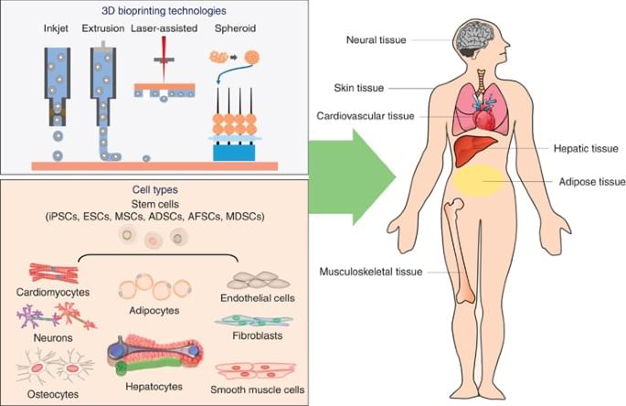
Extrusion-based 3D printing/bioprinting is a promising approach to generating patient-specific, tissue-engineered grafts. However, a major challenge in extrusion-based 3D printing and bioprinting is that most currently used materials lack the versatility to be used in a wide range of applications.

New nanotechnology has been developed by a team of researchers from Texas A&M University that leverages colloidal interactions of nanoparticles to print complex geometries that can mimic tissue and organ structure. The team, led by Dr. Akhilesh Gaharwar, associate professor and Presidential Impact Fellow in the Department of Biomedical Engineering, has introduced colloidal solutions of 2D nanosilicates as a platform technology to print complex structures.

2D nanosilicates are disc-shaped inorganic nanoparticles 20 to 50 nanometers in diameter and 1 to 2 nanometers in thickness. These nanosilicates form a “house-of-cards” structure above a certain concentration in water, known as a colloidal solution.

To begin the process of bioprinting an organ, doctors typically start with a patient’s own cells. They take a small needle biopsy of an organ or do a minimally invasive surgical procedure that removes a small piece of tissue, “less than half the size of a postage stamp,” Atala said. “By taking this small piece of tissue, we are able to tease cells apart (and) we grow and expand the cells outside the body.”

This growth happens inside a sterile incubator or bioreactor, a pressurized stainless steel vessel that helps the cells stay fed with nutrients – called “media” – the doctors feed them every 24 hours, since cells have their own metabolism, Lewis said. Each cell type has a different media, and the incubator or bioreactor acts as an oven-like device mimicking the internal temperature and oxygenation of the human body, Atala said.

“Then we mix it with this gel, which is like a glue,” Atala said. “Every organ in your body has the cells and the glue that holds it together. Basically, that’s also called ‘extracellular matrix.’”.

Fluicell, a bioprinting firm based in Sweden, has launched its new high-precision 3D printer, the Biopixlar AER.

Intended as a successor to the original Biopixlar, the device is Fluicell’s second single-cell 3D bioprinting system. The company has designed its latest machine to be as compact and accessible as possible, and claims that it’s the world’s first microfluidic bioprinter that fits inside a standard flow hood or biosafety cabinet. This enables users to easily integrate it with other in vitro and 3D cell culture technologies.

Victoire Viannay, CEO of Fluicell, said, “With Biopixlar AER, we have reached a new important milestone and we can now offer a pioneering product, fully tailored to meet current and future needs in the rapidly accelerating life science and research sector.”

Medcalf: Because you’re moving away from the economics of scale to closer to the clinic, the batches are smaller and some of the traditional paradigms for quality assurance, such as proof of sterility, are harder to arrange. Thus, you need to have a manufacturing system that includes quality assurance within the system itself.

Automation is often presented as a way to remove the single largest source of infective risk, i.e. the human operator. For example, the self-sterilizing reusable units being developed at the University of Osaka under Professor Masahiro Kino-oka allow small-scale production with a high degree of confidence in the aseptic management of the environment.

Another challenge is defining a product that has variable characteristics. The main reason for decentralizing is to allow customization to a patient, which means you need to have a hierarchy of levels of specification. For example, with bioprinting, which also produces a customized product, you need to define bulk properties, but you also need to set constraints around how it’s anchored or implanted into the patient.

Bioprinting is widely applicable to develop tissue engineering scaffolds and form tissue models in the lab. Materials scientists use this method to construct complex 3D structures based on different polymers and hydrogels; however, relatively low resolution and long fabrication times can result in limited procedures for cell-based applications.

In a new report now available in Nature Asia Materials, Byungjun Lee and a team of scientists in mechanical engineering at Seoul National University, Seoul, Korea, presented a 3D hybrid-micromesh assisted bioprinting method (Hy-MAP) to combine digital light projection, 3D printed micromesh scaffold sutures, together with sequential hydrogel patterning. The new method of bioprinting offered rapid cell co-culture via several methods including injection, dipping and draining. The work can promote the construction of mesoscale complex 3D hydrogel structures across 2D microfluidic channels to 3D channel networks.

Lee et al. established the design rules for Hy-MAP printing via analytical and experimental investigations. The new method can provide an alternative technique to develop mesoscale implantable tissue engineering constructs for organ-on-a-chip applications.

A research team from Utrecht University has successfully fabricated working livers using a newly developed ultrafast volumetric 3D bioprinting method.

By means of visible light tomography, the volumetric bioprinting method enabled the successful printing of miniature stem cell units by making the cells “transparent”, which meant they retained their resolution and ability to perform biological processes.

Printed in less than 20 seconds, the liver units were able to perform key toxin elimination processes mimicking those that natural livers perform in our bodies, and could open new opportunities for regenerative medicine and personalized drug testing.

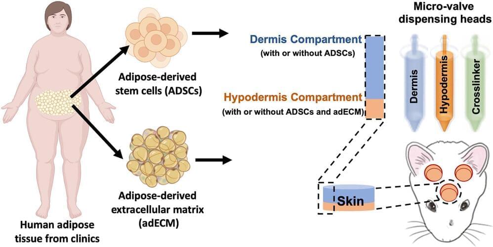
Researchers from the University of Birmingham and the University of Huddersfield, UK, have developed a new 3D bioprinting technique that can be used to treat chronic wounds.

Named Suspended Layer Additive Manufacturing (SLAM), the approach enables the printing of a novel biomaterial that accurately simulates the structure of mammalian skin.

In fact, according to the researchers, the biomaterial is the first of its kind to simulate all three of the major layers found in skin – the hypodermis, the dermis, and the epidermis – making it a unique tri-layered skin equivalent. Early experiments suggest that the 3D bioprinted skin can be placed at the site of a wound to induce healing, reducing scar tissue in the process.