Toggle light / dark theme

Bioengineers build branched, perfusable kidney collecting ducts using 3D bioprinting

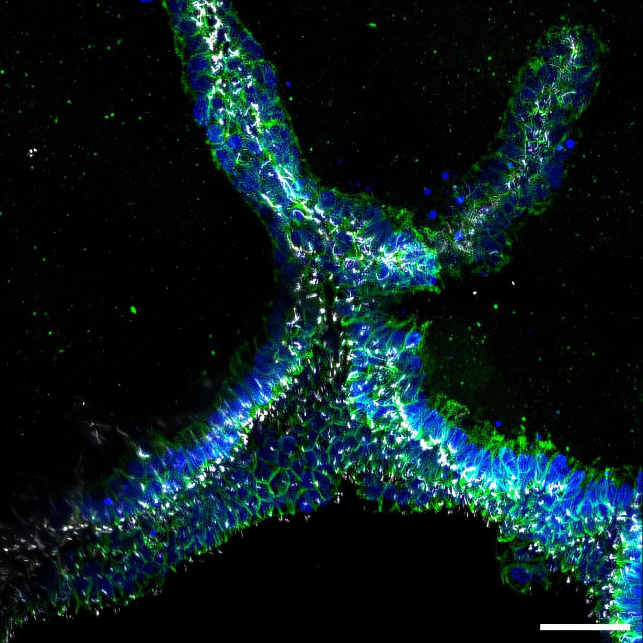
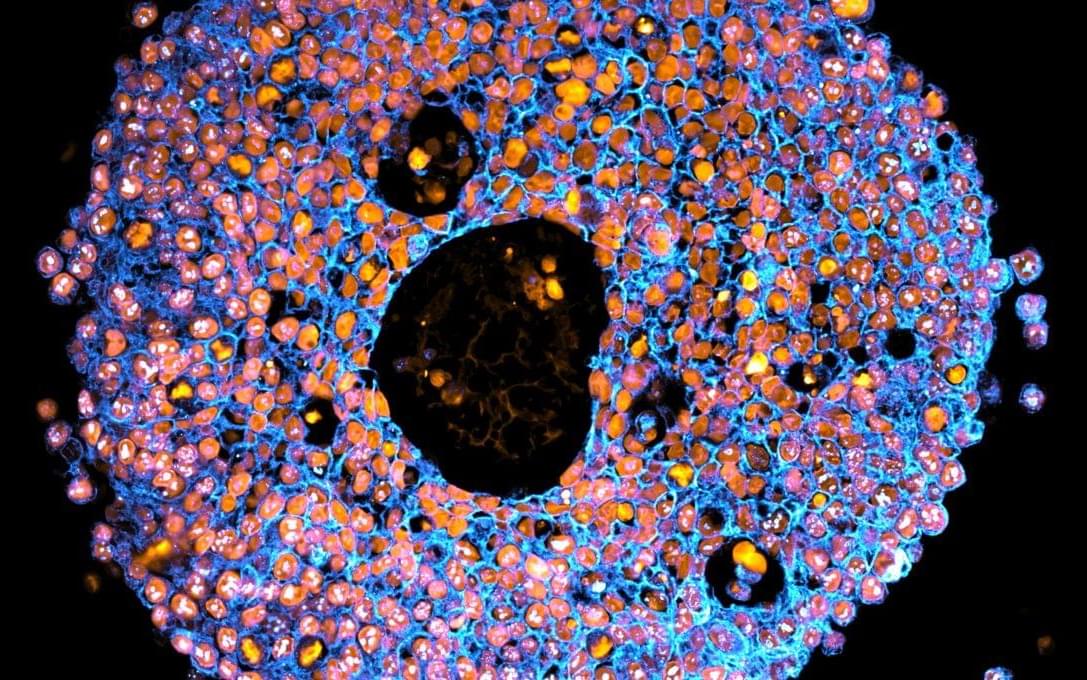
The human kidney filters about a cup of blood every minute, removing waste, excess fluid, and toxins from it, while also regulating blood pressure, balancing important electrolytes, activating Vitamin D, and helping the body produce red blood cells. This broad range of functions is achieved in part via the kidney’s complex organization. In its outer region, more than a million microscopic units, known as nephrons, filter blood, reabsorb necessary nutrients, and secrete waste in the form of urine.

To direct urine produced by this enormous number of blood-filtering units to a single ureter, the kidney establishes a highly branched three-dimensional, tree-like system of “collecting ducts” during its development. In addition to directing urine flow to the ureter and ultimately out of the kidney, collecting ducts reabsorb water that the body needs to retain, and maintain, the body’s balance of salts and acidity at healthy levels.

Finding ways to recreate this system of collecting ducts is the focus of researchers and bioengineers who are interested in understanding how duct defects cause certain kidney diseases, underdeveloped kidneys, or even the complete absence of a kidney. Being able to fabricate the kidney’s plumbing system from the bottom up would be a giant step toward tissue replacement therapies for many patients waiting for a kidney donation: In the U.S. alone, 90,000 patients are on the kidney transplant waiting list. However, rebuilding this highly branched fluid-transporting ductal system is a formidable challenge and not possible yet.

Nanoengineered extrusion-aligned tract bioprinting enables functional repair of spinal cord injuries

Gu et al. present NEAT, a nanoengineered extrusion-aligned tract bioprinting strategy that fabricates aligned, human neural stem cell-laden collagen hydrogel constructs through shear-induced fibrillar organization. In a rat model of complete spinal cord transection, NEAT enables axonal reconnection and functional locomotor recovery, demonstrating its translational potential for spinal cord repair and neural tissue engineering.

Programmable Macrophage Mimics for Inflammatory Meniscus Regeneration via Nanotherapy

JUST PUBLISHED: programmable macrophage mimics for inflammatory meniscus regeneration via nanotherapy

Click here to read the latest free, Open Access Article from Research.


The meniscus is a fibrocartilaginous tissue and organ in the human knee joint that serves critical functions, including load transmission, shock absorption, joint stability, and lubrication. Meniscal injuries are among the most common knee injuries, typically caused by acute trauma or age-related degeneration [13]. Minor meniscal injuries are usually treated with in situ arthroscopic procedures or conservative methods, whereas larger or more severe injuries often necessitate total meniscus replacement. Recent advances in materials science and manufacturing techniques have enabled transformative tissue-engineering strategies for meniscal therapy [4, 5]. Several stem cell types, including synovium-derived mesenchymal stem cells, bone-marrow-derived mesenchymal stem cells, and adipose-derived stem cells (ADSCs), have been investigated as candidate seed cells for meniscal regeneration and repair. Notably, ADSCs are clinically promising because of their ease of harvest, high inducibility, innate anti-inflammatory properties, and potential to promote fibrocartilage regeneration [68]. Our group has developed a series of decellularized matrix scaffolds for auricular, nasal, tracheal, and articular cartilage repair using 3-dimensional (3D) bioprinting techniques, successfully repairing meniscus defects and restoring physiological function [912]. However, current tissue-engineering strategies for meniscus defect repair commonly rely on a favorable regenerative microenvironment. Pathological conditions such as osteoarthritis (OA) [13 16], the most prevalent joint disorder, often create inflammatory environments that severely hinder meniscus regeneration [17 21]. Moreover, meniscal injury exacerbates the local inflammatory milieu, further impeding tissue healing and inevitably accelerating OA progression. Therefore, there is an urgent need to establish a cartilaginous immune microenvironment that first mitigates early-stage inflammation after meniscal injury and then sequentially promotes later-stage fibrocartilage regeneration [22 25].

Currently, targeted regulation using small-molecule drug injections is commonly employed to treat inflammatory conditions in sports medicine [26,27]. Most of these drugs exhibit broad-spectrum anti-inflammatory effects and inevitably cause varying degrees of side effects by activating nonspecific signaling pathways. Polyethyleneimine is a highly cationic polymer. It is widely used to modulate inflammation by adsorbing and removing negatively charged proinflammatory cytokines, including tumor necrosis factor-α (TNF-α), interleukin-1β (IL-1β), and interleukin-6 (IL-6), via electrostatic interactions [28–31]. Notably, modifying polyethyleneimine into its branched form (branched polyethyleneimine [BPEI]) has been shown to improve cytocompatibility and enhance in vivo metabolic cycling.

The Incredible Science of Bioprinting

Dive into the remarkable world of bioprinting in this comprehensive video. We’ll be exploring the core concepts of bioprinting — a pioneering technique that uses biological materials to create structures that mimic natural tissues, organs, and even cells. Understand the sophisticated science behind this process, and learn how bio-inks are formulated and layered to build live cells. We’ll also embark on a historical journey, tracing the origins and evolution of bioprinting, and how it is reshaping modern medicine. From overcoming organ shortages to paving the way for personalized treatments, bioprinting is revolutionizing healthcare. Join us as we unpack this fascinating technology and its promising future.

For more information on how patents can help drive your innovation.
visit https://www.patsnap.com/

From concept to commercialization, Patsnap’s innovation intelligence platform breaks boundaries to connect innovators with insights.

Follow us on social for more!
LinkedIn: / patsnap.
Facebook: / patsnap.
Twitter: / patsnap.
Instagram: / _patsnap.

PatSnap (UK) Limited (PatSnap) hereby disclaims all representations and warranties, express or implied, with respect to information, advertisements and all other ad-hoc materials published by us. PatSnap does not represent or warrant that the information accessible on this material is accurate, complete or current, and makes no claims or warranties of any kind, including the suitability of information, or merchantability and fitness for a particular purpose. It is the sole responsibility of the Recipient of such information to comply with all applicable laws, rules and regulations and they are responsible for meeting any regulatory, compliance and legal obligations. All due-diligence and vetting of services are the sole responsibility of the Recipient.

Revitalizing liver function in mice with liver failure through transplantation of 3D-bioprinted liver with expanded primary hepatocytes

Year 2024 face_with_colon_three


3D-bioprinting livers based on expandable primary hepatocytes and liver-specific bioink could save mouse liver failure.

Droplet-based bioprinting Reviews Methods Primers

(DBB) enables the fabrication of three-dimensional structures from biomaterials. In this Primer, Gupta and colleagues describe the various DBB modalities and their applications, considerations for the selection of bioinks and substrates, methods to assess printability, the limitations of DBB and their solutions, and potential advances that might be brought about by incorporating new technologies.

A pill that prints bio-ink for damaged tissue repair

EPFL researchers have demonstrated the first pill-sized bioprinter that can be swallowed and guided within the gastrointestinal tract, where it directly deposits bio-ink over damaged tissues to support repair.

Soft tissue injuries of the , like ulcers or hemorrhages, can currently be treated only with some form of surgery, which is invasive and may not result in permanent repair. Bioprinting is emerging as an effective treatment that deposits biocompatible “ink”—often made of natural polymers derived from seaweed—directly over the site of tissue damage, creating a scaffold for new cell growth. But like traditional surgical tools, these kinds of bioprinters tend to be bulky and require anesthesia.

At the same time, “untethered” technologies are being developed to perform medical interventions without a physical connection to external equipment. For example, ingestible “smart capsules” can be guided to drug delivery sites using external magnets. But these devices are designed to travel through liquids, and their movements become unpredictable when they touch the tissue wall.

Breakthrough 3D Bioprinted Mini Placentas May Help Solve “One Of Medicine’s Great Mysteries”

To address these shortcomings, the team behind the latest study turned to bioprinting – a type of 3D printing that uses living cells and cell-friendly materials to create 3D structures. They took trophoblast cells and mixed them with a synthetic gel before 3D-printing them in precise droplets.

The printed cells then grew into miniature placentas, and the researchers compared them to organoids made via traditional manual methods.

“The organoids we grew in the bioprinted gel developed differently to those grown in an animal-derived gel, and formed different numbers of trophoblast sub-types. This highlighted that the environment organoids are grown in can control how they mature,” first author Dr Claire Richards said.

/* */