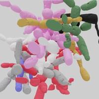
Evolution’s rapid pace after the Cambrian explosion
Though the work of Schopf and other paleobiologists continues to fill in the Precambrian fossil record, questions remain about the pace of the Cambrian explosion. What triggered life to evolve so fast?
The question has intrigued scientists of many disciplines for decades. Interdisciplinary collaboration has wrought a wealth of evidence from diverse perspectives — geochemical, paleoenvironmental, geological, anatomical, and taxonomic — that describes how biological organisms evolved in concert with changing environmental conditions.
Keep exploring at http://brilliant.org/ArtemKirsanov/ Get started for free, and hurry—the first 200 people get 20% off an annual premium subscription.
My name is Artem, I’m a computational neuroscience student and researcher. In this video we will see why individual neurons essentially function like deep convolutional neural networks, equipped with insane information processing capabilities as well as some of the physiological mechanisms, that account for such computational complexity.
OUTLINE: 00:00 Introduction. 01:42 — Perceptrons. 03:43 — Electrical excitability and action potential. 07:12 — Cable theory: passive dendrites. 09:03 — Active dendritic properties. 12:10 — Human neurons as XOR gates. 19:11 — Single neurons as deep neural networks. 22:32 — Brilliant. 23:57 — Recap and outro.
REFERENCES (in no particular order): 1. Bicknell, B. A., Bicknell, B. A. & Häusser, M. A synaptic learning rule for exploiting nonlinear dendritic computation. Neuron (2021) doi:10.1016/j.neuron.2021.09.044. 2. Matthew Larkum. Are dendrites conceptually useful? Neuroscience (2022) doi:10.1016/j.neuroscience.2022.03.008. 3. Polsky, A., Mel, B. W. & Schiller, J. Computational subunits in thin dendrites of pyramidal cells. Nature Neuroscience 7621–627 (2004). 4. Tran-Van-Minh, A. et al. Contribution of sublinear and supralinear dendritic integration to neuronal computations. Frontiers in Cellular Neuroscience 9, 67–67 (2015). 5. Gidon, A. et al. Dendritic action potentials and computation in human layer 2/3 cortical neurons. Science 367, 83–87 (2020). 6. London, M. & Häusser, M. DENDRITIC COMPUTATION. Annu. Rev. Neurosci. 28503–532 (2005). 7. Branco, T., Clark, B. A. & Häusser, M. Dendritic Discrimination of Temporal Input Sequences in Cortical Neurons. Science 329, 1671–1675 (2010). 8. Stuart, G. J. & Spruston, N. Dendritic integration: 60 years of progress. Nat Neurosci 18, 1713–1721 (2015). 9. Smith, S. L., Smith, I. T., Branco, T. & Häusser, M. Dendritic spikes enhance stimulus selectivity in cortical neurons in vivo. Nature 503115–120 (2013). 10. Beniaguev, D., Segev, I. & London, M. Single cortical neurons as deep artificial neural networks. Neuron 109, (2021). 11. Michalikova, M., Remme, M. W. H., Schmitz, D., Schreiber, S. & Kempter, R. Spikelets in pyramidal neurons: generating mechanisms, distinguishing properties, and functional implications. Reviews in the Neurosciences 31101–119 (2019). 12. Larkum, M. E., Wu, J., Duverdin, S. A. & Gidon, A. The Guide to Dendritic Spikes of the Mammalian Cortex In Vitro and In Vivo. Neuroscience 489, 15–33 (2022).
How do plants defend themselves against pathogenic microorganisms? This is a complex puzzle, of which a team of biologists from the University of Amsterdam has solved a new piece. The team, led by Harrold van den Burg, discovered that while the water pores (hydathodes) in leaves provide an entry point for bacteria, they are also an active part of the defense against these invaders. The team’s research has now been published in the journal Current Biology.
Anyone who is used to giving plants plenty of water might know the phenomenon: small droplets of plant sap that sometimes appear at the edge of the leaves, especially at nighttime. When plants take up more water via their roots than they lose through evaporation, they can use their water pores on the leaf margins to release excess water. The pores literally prevent root water pressure from becoming too high. This is an important mechanism, but at the same time, risky. Pathogenic microorganisms can enter the plant’s veins through these sap droplets to colonize the water pores.
Biologists have therefore been asking themselves for a long time: How do plants defend themselves against this wide-open entry point? Are those water pores, the hydathodes, defenseless glands that allow ample entry of harmful pests? Or have they evolved in such a way that they are part of the plant’s line of defense against pathogens?
A team of researchers from Illinois Institute of Technology and the University of Washington is trying to change the way that the field of biology understands how muscles contract.
In a paper published on January 25, 2023, in the Proceedings of the National Academy of Sciences titled “Structural OFF/ON Transition of Myosin in Related Porcine Myocardium Predict Calcium Activated Force,” Illinois Tech Research Assistant Professor Weikang Ma and Professor of Biology and Physics Thomas Irving—working in collaboration with Professor of Bioengineering Michael Regnier’s group at Washington—make the case for a second, newly discovered aspect to muscle contraction that could play a significant role in developing treatments for inherited cardiac conditions.
The consensus for how muscle contraction occurs has been that the relationship between the thin and thick filaments that comprise muscle tissue was a more straightforward process. When targets on thin filaments were activated, it was thought that the myosin motor proteins that make up the thick filaments would automatically find their way to those thin filaments to start generating force and contract the muscle.
Artificial intelligence has already shaved years off research into protein engineering. Now, for the first time, scientists have synthesized proteins predicted by an AI model in the lab, and found them to work just as well as their natural counterparts.
The first time a language model was used to synthesize human proteins.
Of late, AI models are really flexing their muscles. We have recently seen how ChatGPT has become a poster child for platforms that comprehend human languages. Now a team of researchers has tested a language model to create amino acid sequences, showcasing abilities to replicate human biology and evolution.
The language model, which is named ProGen, is capable of generating protein sequences with a certain degree of control. The result was achieved by training the model to learn the composition of proteins. The experiment marks the first time a language model was used to synthesize human proteins.
Blueprint is a public science experiment to determine whether it’s possible to stay the same biological age. This requires slowing down aging processes as much as possible and then reversing the aging that has happened. Currently my speed of aging is .76 (DunedinPACE). That means for every 365 days each year, I age 277 days. My goal is to remain the same age biologically for every 365 days that pass.
I openly share (for free!) my diet, exercise and other protocols so that others can benefit and try to improve upon what I’m doing. I also openly share my health data as data is better than human opinion at guiding decision making. You can find everything here: https://blueprint.bryanjohnson.co/
This video is a whirlwind tour of what my daily life looks like as my team and I are on this adventure.
On the surface, Blueprint may seem like something about health, wellness and aging. To me, it’s a philosophy.
Dr. Bapteste has both a Ph.D. in evolutionary biology from Pierre and Marie Curie University and a Ph.D. in the philosophy of biology from Pantheon-Sorbonne University.
Dr. Bapteste is the Co-Director of the Adaptation, Intégration, Réticulation, Evolution (AIRE) team, which develops new methods and new concepts, in particular related to biological networks, in order to study evolution and aging. Specifically, the AIRE team works to enhance the evolutionary theory i) by expanding its scope by targeting additional objects of studies (such as novel units of selection and novel still unknown taxonomical groups from the microbial dark matter, and mobile elements) and ii) by expanding evolutionary studies towards more general models, able to in particular account for chimerism and interactions between biological elements, from molecules to ecosystems.
Dr. Bapteste is the author of 95 scientific articles and 4 books of popular sciences: “Les gènes voyageurs: l’odyssée de l’évolution”, “Conflits intérieurs: fable scientifique”, “Tous entrelacés! Des gènes aux super-organismes, les réseaux de l’évolution”, and “Tout se transforme! Comment marche l’évolution”.
What led to the emergence of complex organisms on Earth? It’s a significant unanswered question in biology. Researchers from Christa Schleper’s team at the University of Vienna and Martin Pilhofer’s team at ETH Zurich have taken a step towards resolving it. The scientists succeeded in cultivating a special archaeon and characterizing it more precisely using microscopic methods.
This member of the Asgard archaea exhibits unique cellular characteristics and may represent an evolutionary “missing link” to more complex life forms such as animals and plants. The study was recently published in the journal Nature.
All life forms on earth are divided into three major domains: eukaryotes, bacteria and archaea. Eukaryotes include the groups of animals, plants and fungi. Their cells are usually much larger and, at first glance, more complex than the cells of bacteria and archaea. The genetic material of eukaryotes, for example, is packaged in a cell nucleus and the cells also have a large number of other compartments. Cell shape and transport within the eukaryotic cell are also based on an extensive cytoskeleton. But how did the evolutionary leap to such complex eukaryotic cells come about?
A model for information storage in the brain reveals how memories decay with age.
Theoretical constructs called attractor networks provide a model for memory in the brain. A new study of such networks traces the route by which memories are stored and ultimately forgotten [1]. The mathematical model and simulations show that, as they age, memories recorded in patterns of neural activity become chaotic—impossible to predict—before disintegrating into random noise. Whether this behavior occurs in real brains remains to be seen, but the researchers propose looking for it by monitoring how neural activity changes over time in memory-retrieval tasks.
Memories in both artificial and biological neural networks are stored and retrieved as patterns in the way signals are passed among many nodes (neurons) in a network. In an artificial neural network, each node’s output value at any time is determined by the inputs it receives from the other nodes to which it’s connected. Analogously, the likelihood of a biological neuron “firing” (sending out an electrical pulse), as well as the frequency of firing, depends on its inputs. In another analogy with neurons, the links between nodes, which represent synapses, have “weights” that can amplify or reduce the signals they transmit. The weight of a given link is determined by the degree of synchronization of the two nodes that it connects and may be altered as new memories are stored.