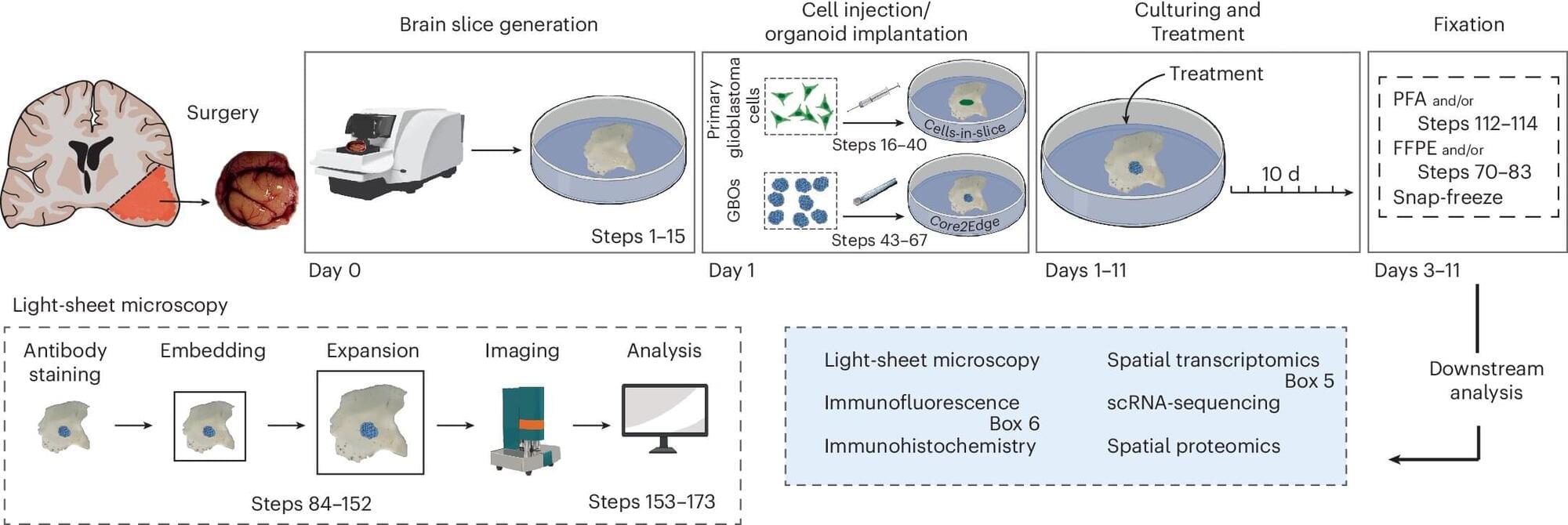
Toggle light / dark theme

Get the latest international news and world events from around the world.

Log in for authorized contributors

Parkinson Disease Pathogenic VariantsCross-Ancestry Analysis and Microarray Data Validation

Parkinson disease pathogenic variants: cross-ancestry analysis and microarray data validation.


This website uses a security service to protect against malicious bots. This page is displayed while the website verifies you are not a bot.

Immune molecule long tied to inflammation may benefit the aging brain

Inflammation in the brain is usually seen as harmful in the aging process—it’s thought to contribute to Alzheimer’s and dementia. But a new study in mice suggests that inflammation, led by an immune molecule called STING (stimulator of interferon genes), might have a role in protecting the aging brain. The findings also have implications for new experimental Alzheimer’s drugs that are designed to block STING.

For the study published in Cell Reports, scientists at Tufts University School of Medicine examined brain function, inflammation, and movement in genetically engineered to lack STING, compared with normal controls. They found that mice without STING had worse memory and movement problems, mimicking the senility and frailty seen in people with dementia and Alzheimer’s disease.

“Our data suggest that the that STING supports may actually be necessary for the brain to stay healthy and in balance during old age,” says Shruti Sharma, an assistant professor of immunology at Tufts University School of Medicine and the study’s senior author.

HES1 regulates bone marrow mesenchymal stromal cell function by suppressing NFATc2-mediated inflammation

The bone marrow microenvironment plays a central role in hematopoiesis and hematologic diseases. A new study investigates how HES1 regulates mesenchymal stromal cell function and inflammatory signaling, exploring a novel pathway that may shape hematopoietic homeostasis and disease biology.


The Notch target gene, Hairy and enhancer of split-1 (HES1), encodes a basic helix-loop-helix transcriptional repressor that influences cell proliferation and differentiation in embryogenesis. Our previous studies indicate that HES1 is required for hematopoiesis under stress conditions. However, the role of HES1 in bone marrow (BM) microenvironment remains to be elucidated. By employing a BM niche specific Hes1 knockout mouse model, here we have investigated the role of HES1 in regulating mesenchymal stromal cell (MSC) homeostasis and their hematopoiesis supportive function. We found that while HES1 is not essential in MSC in supporting steady-state hematopoiesis, Hes1fl/fl Prx1Cre mice are hypersensitive to lipopolysaccharide (LPS) challenge. Deletion of Hes1 in the BM reduces MSC frequency and affects MSC self-renewal and proliferation. Hes1-deficient MSC are less functional in supporting hematopoiesis both in vitro and ex vivo. Transcriptome analysis reveals that disruption of Hes1 in the BM stroma alters the expression of genes critical for cellular metabolism and inflammation. Pharmacological blockage of inflammation rescues Hes1-KO MSC phenotype and improves their hematopoiesis supportive function. Mechanistically, we show that HES1 binds to the conserved E boxes in the promoter of NFATc2, a member of the AT-rich interaction domain superfamily of DNA binding protein, to suppress NFATc2-mediated inflammation. Taken together, our study unveils a pivotal role for HES1 in maintaining BM MSC hemostasis and regulating their hematopoiesis supportive function.

Hematopoietic stem cells (HSC), which give rise to all blood cells, are supported by specialized microenvironments, known as niches, within bone marrow (BM) cavities.1,2 These niches are composed of various non-hematopoietic components, including endosteal and sinusoidal endothelial cells, mesenchymal stromal cells (MSC), and osteoblast-lineage cells.3,4 Studies have shown that BM-derived non-hematopoietic stromal cells are capable of supporting long-term hematopoiesis both in vitro and in vivo. Disruptions to these non-hematopoietic cells within the BM niche may negatively impact hematopoiesis. However, the precise mechanism is still poorly understood.

Notch signaling, mediated by a family of highly conserved receptors, plays a critical role in maintaining bone homeostasis, partly through regulating osteoblast differentiation from MSC, whose activation is induced by their specific ligands.5,6

Mirtazapine for Methamphetamine Use Disorder: A Randomized Clinical Trial

In adults with methamphetamine use disorder, mirtazapine reduced methamphetamine use by approximately two more days per month vs placebo, with no unexpected safety concerns.


Main Outcomes and Measures The primary end point was the change in days of methamphetamine use in the past 28 days from baseline to week 12. Secondary end points were depression, insomnia, HIV risk behavior, quality of life, and methamphetamine-negative oral fluid samples.

Results Of 344 participants randomized, 339 participants received the intervention (167 in the placebo group and 172 in the mirtazapine group). Mean (SD) age was 42.0 (8.6) years, 126 participants (37.2%) were female, and participants had used methamphetamine for a median (IQR) of 24 days (17−28) of the past 28 days at baseline. The mean reduction in days of methamphetamine use from baseline to week 12 was greater in the mirtazapine group (7.0 days of 28 days) than in the placebo group (4.8 days of 28 days; mean difference, 2.2 days; 95% CI, −4.2 to −0.2 days; P = .02). More participants in the mirtazapine group reported drowsiness (47% vs 33%) and weight gain (10% vs 3%). Forty participants (23%) discontinued mirtazapine due to adverse events compared to 25 participants (15%) in the placebo group. No significant effects of mirtazapine on secondary end points were found.

Conclusions and Relevance In this parallel-group randomized clinical trial, mirtazapine delivered in routine clinical practice reduced methamphetamine use in adults with methamphetamine use disorder. No unexpected safety concerns delivering mirtazapine in this setting were found; this finding has important clinical implications in the absence of any approved pharmacotherapies for methamphetamine use disorder.

Your brain may be as blind to reality as a grasshopper is to calculus | Michelle Thaller

In the distant future could we redesign the brain for more understanding of the universe.


Become a Big Think member to unlock expert classes, premium print issues, exclusive events and more: https://bigthink.com/membership/?utm_… What if the universe was never designed to be understood? Astronomer Michelle Thaller makes a case that the human brain, despite its complexity, may be as poorly equipped to grasp ultimate reality as a grasshopper is to.

What if the universe was never designed to be understood? Astronomer Michelle Thaller makes a case that the human brain, despite its complexity, may be as poorly equipped to grasp ultimate reality as a grasshopper is to grasp calculus.

About Michelle Thaller:

Michelle Thaller is an astronomer and Assistant Director for Science Communication at NASA’s Goddard Space Flight Center.

The frequency-dependent effects of primary hand motor cortex stimulation on volitional finger movement

[Brain stimulation] Taquet et al.: “The effect of motor cortex stimulation on finger flexion is frequency dependent.”


We conducted a prospective study in human patients undergoing awake craniotomies to examine whether the effects of cortical stimulation in hand primary motor cortex (M1) can be frequency dependent and inhibitory.

In 11 participants undergoing clinically indicated awake craniotomies, we delivered bursts of 1–333 Hz stimulation during a finger-flexion task. Synchronized electrocorticography (ECoG), finger joint kinematics, electromyography (EMG), and video were recorded.

Inability to flex the index finger during subthreshold stimulation was noted in 3 participants at frequencies 250 Hz when the electrodes were in locations that induced extension of the forefinger at higher amplitudes. Other than these trials, all stimulation events either induced muscle contractions or had no measurable effect.

Programmed Cell Death Protein 1 (PD-1) and Programmed Cell Death Ligand 1 (PD-L1) Immunotherapy: A Promising Breakthrough in Cancer Therapeutics

PD-1/PD-L1 Inhibitors Implications in Common Human Cancers.

Lung cancer: the landscape of lung cancer treatment has been profoundly reshaped by tumor immunotherapy directed at PD-1/PD-L1. Notably, the effectiveness of PD-L1 inhibitors surpasses that of chemotherapy, particularly in advanced non-small cell lung cancer (NSCLC) patients exhibiting elevated PD-L1 levels. This potency is equally evident among patients with previously untreated metastatic squamous NSCLC. Moreover, when considering patients with NSCLC who have undergone prior treatment, a decreased rate of disease progression is more frequently observed in response to PD-1/PD-L1 inhibitors, as opposed to conventional chemotherapy. This observation holds true, particularly for patients with an extensive metastatic burden and an adverse prognosis. In current clinical therapeutics, a strategic alliance between PD-1/PD-L1 immune checkpoint inhibitors and chemotherapeutic agents has emerged as a cornerstone of treatment. This approach attests to the heightened value these inhibitors bring to the therapeutic arsenal. The rapid evolution of anti-PD-1/PD-L1 inhibitors for advanced NSCLC stands as an instrumental factor in enhancing patient outcomes, charting a promising trajectory toward improved prognosis [,]. In a recent study, neoadjuvant PD-1 inhibitor sintilimab was administered to individuals with NSCLC. The outcomes revealed that a notable 40.5% of participants achieved a major pathological response, while a commendable 10.8% realized a complete remission at the pathological level [].

Prostate cancer: currently, PD-1/PD-L1 immune checkpoint inhibitors have ushered substantial clinical advantages for individuals with prostate cancer. A recent study has put forth the notion that synergizing PD-1/PD-L1 checkpoint inhibitors with radiotherapy presents a promising avenue in the management of prostate cancer []. However, it is noteworthy that the impact of PD-L1/PD-1 blockade in the context of prostate cancer appears comparatively muted in contrast to its influence on other cancer types. This discrepancy stems from the diminished immunogenicity characterizing prostate cancers [].

First Signs of Quark–Gluon Plasma in Oxygen–Oxygen Collisions

When two heavy nuclei collide at relativistic speeds, the quarks and gluons that are usually bound inside them are briefly liberated, forming an exotic state of matter called quark–gluon plasma. As the quarks and gluons traverse this plasma, they lose energy through scattering, which limits the number of high-momentum particles that reach the detectors. This signature of quark–gluon plasma, called jet quenching, has been definitively observed only in collisions of heavy nuclei such as lead, leaving open the question of how large a nucleus must be to produce quark–gluon plasma. Now the CMS Collaboration at the Large Hadron Collider (LHC) at CERN has observed the first clear evidence of jet quenching in oxygen–oxygen collisions [1].

The LHC collided oxygen nuclei for the first time in 2025. Scientists in the CMS Collaboration measured the rate at which those collisions produced high-momentum daughter particles and compared it to the rate measured for proton–proton collisions at the same collision energy. In the absence of a quark–gluon plasma, the two rates—after accounting for the number of protons and neutrons in the oxygen nuclei—would be approximately equal. The researchers found that, in the oxygen–oxygen collisions, this ratio dipped significantly for daughter particles with energies of around 6 giga-electron-volts (GeV)—a clear indication of the jet-quenching phenomenon.

The oxygen–oxygen collision data recorded by the CMS team are qualitatively similar to those obtained from collisions of larger nuclei such as lead. They are also in better agreement with theoretical models that include quark–gluon energy loss than they are with models that omit it. The result provides the strongest evidence yet that a quark–gluon-plasma-like medium capable of jet quenching can form in collisions of nuclei as light as oxygen.

For 74,000 years, one ancient killer quietly dictated where early humans could survive across Africa

Increasing evidence suggests that our species emerged through interactions between populations living in different parts of Africa, rather than from a single birthplace. Until now, however, most explanations for how those populations were distributed across the continent have focused on climate alone. The new research shows that disease—specifically malaria—also played a crucial role.

In a paper published in Science Advances, researchers from the Max Planck Institute of Geoanthropology, the University of Cambridge, and colleagues have investigated whether Plasmodium falciparum-induced malaria shaped human habitat choice between 74,000 and 5,000 years ago, the critical period before humans dispersed widely beyond Africa and before agriculture dramatically altered malaria transmission.

The study shows that malaria, one of humanity’s oldest and most persistent pathogens, influenced habitat choice by pushing human groups away from high-risk environments and separating populations across the landscape. Over tens of thousands of years, this fragmentation shaped how populations met, mixed, and exchanged genes, helping create the population structure seen in humans today. The findings suggest that infectious disease was not simply a challenge early humans faced: it was a fundamental factor shaping the deep history of our species.

/* */