Toggle light / dark theme

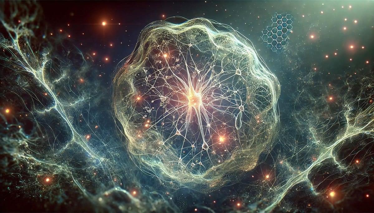
This essay advances a speculative yet empirically-grounded hypothesis: that microtubular cytoskeletal structures constitute proto-cognitive architectures in unicellular organisms, thereby establishing an evolutionary substrate for cognition that predates neural systems. Drawing upon converging evidence from molecular biology, quantum biophysics, phenomenological philosophy, and biosemiotic theory, I propose a cytoskeletal epistemology wherein cognition emerges not exclusively from neural networks, but from the dynamic, embodied information-processing capacities inherent in cellular organization itself. This framework challenges neurocentric accounts of mind while suggesting new avenues for investigating the biological foundations of knowing.

Contemporary cognitive science predominantly situates the genesis of mind within neural tissue, tacitly assuming that cognition emerges exclusively from the electrochemical dynamics of neurons and their synaptic interconnections. Yet this neurocentric paradigm, while experimentally productive, encounters both conceptual and empirical limitations when confronted with fundamental questions regarding the biological preconditions for epistemic capacities. As Thompson (2007) observes, “Life and mind share a set of basic organizational properties, and the organizational properties distinctive of mind are an enriched version of those fundamental to life” (p. 128). This suggests a profound continuity between biological and cognitive processes — a continuity that invites investigation into pre-neural substrates of cognition.

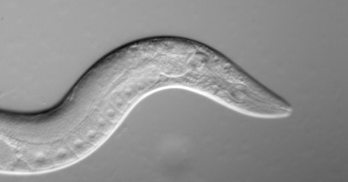
The present inquiry examines the hypothesis that the microtubule — a foundational cytoskeletal element ubiquitous across eukaryotic cells — functions not merely as mechanical infrastructure but as an evolutionary precursor to cognitive architecture, instantiating proto-epistemic capacities in unicellular and pre-neural multicellular organisms. This hypothesis emerges at the intersection of multiple research programs, including quantum approaches to consciousness (Hameroff & Penrose, 2014), autopoietic theories of cognition (Maturana & Varela, 1980), and recent advances in cytoskeletal biology (Pirino et al., 2022).

Miniature zombies are all around us, scuttling through the underbrush or flying through the air in nearly every continent on Earth. In Brazil, a fungus takes over ant brains, altering their circadian rhythms and social behaviors. In England, a virus forces caterpillars to climb high into the canopy, then slowly liquefies their bodies, which drip onto the leaves below. In Indonesia, a parasitoid wasp uses specialized venom to alter a cockroach’s brain chemistry, turning it into the perfect host for her young.

In her new book, Rise of the Zombie Bugs, self-described professional science nerd Mindy Weisberger introduces readers to a menagerie of mind-controlling parasites, and the scientists who have devoted their lives to the study of these peculiar organisms. Through these vivid tales of creatures bizarre enough to rival any fictional beast, Weisberger offers readers a peek into the fields of evolution, ecology, neuroscience, and molecular biology. She shows that these topics exist beyond dim lecture halls and dry textbooks: “Science is everything and everywhere,” she said.

Until now, Google’s Android XR glasses had only appeared in carefully curated teaser videos and limited hands-on previews shared with select publications. These early glimpses hinted at the potential of integrating artificial intelligence into everyday eyewear but left lingering questions about real-world performance. That changed when Shahram Izadi, Google’s Android XR lead, took the TED stage – joined by Nishtha Bhatia – to demonstrate the prototype glasses in action.

The live demo showcased a range of features that distinguish these glasses from previous smart eyewear attempts. At first glance, the device resembles an ordinary pair of glasses. However, it’s packed with advanced technology, including a miniaturized camera, microphones, speakers, and a high-resolution color display embedded directly into the lens.

The glasses are designed to be lightweight and discreet, with support for prescription lenses. They can also connect to a smartphone to leverage its processing power and access a broader range of apps.

Researchers from Tokyo Metropolitan University have found that the motion of unlabeled cells can be used to tell whether they are cancerous or healthy. They observed malignant fibrosarcoma cells and healthy fibroblasts on a dish and found that tracking and analysis of their paths can be used to differentiate them with up to 94% accuracy.

Beyond diagnosis, their technique may also shed light on -related functions, like tissue healing. The paper is published in the journal PLOS ONE.

While scientists and medical experts have been looking at cells under the microscope for many centuries, most studies and diagnoses focus on their shape, what they contain, and where different parts are located inside. But cells are dynamic, changing over time, and are known to be able to move.

Vision is one of the most crucial human senses, yet more than 300 million people worldwide are at risk of vision loss due to various retinal diseases. While recent advancements in retinal disease treatments have successfully slowed disease progression, no effective therapy has been developed to restore already lost vision—until now.

KAIST researchers led by Professor Jinwoo Kim from the Department of Biological Sciences have successfully developed a novel drug to restore vision through retinal nerve regeneration. The research is published in the journal Nature Communications. The study was co-authored by Dr. Eun Jung Lee of Celliaz Inc. and Museong Kim, a Ph.D. candidate at KAIST, as joint first authors.

The research team successfully induced neural regeneration and vision recovery in a disease-model mouse by administering a compound that blocks the PROX1 (Prospero Homeobox 1) protein, which suppresses retinal regeneration. The effect lasted for more than six months.