Toggle light / dark theme

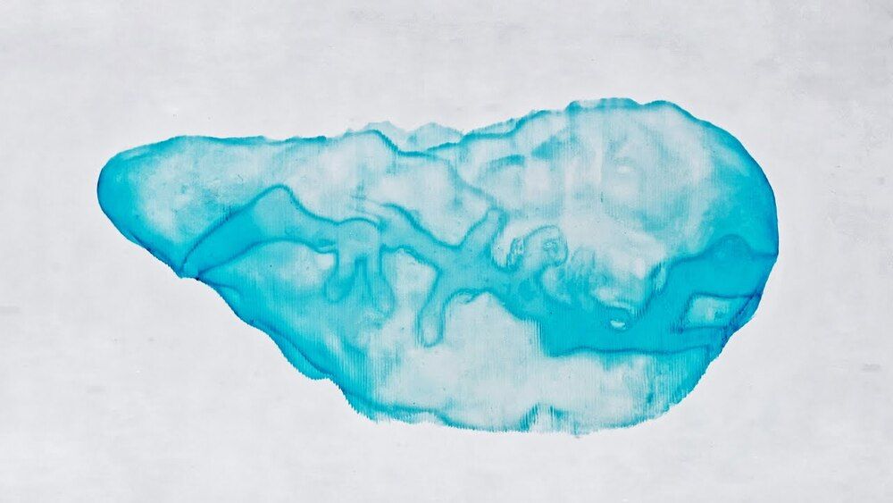
Volumetric 3D bioprinter manufacturer and EPFL spin-out Readily3D has taken the first step towards developing a 3D printed living model of the human pancreas for testing diabetes medicines.

Readily3D’s novel technology is being deployed within the EU-funded Enlight project and is reportedly capable of 3D printing a biological tissue containing human stem cells in just 30 seconds.

As the project’s official bioprinter manufacturer, the company has adapted its proprietary contactless tomographic illumination technology to suit the specific needs of pancreatic tissue structures.

Circa 2019 o,.o.


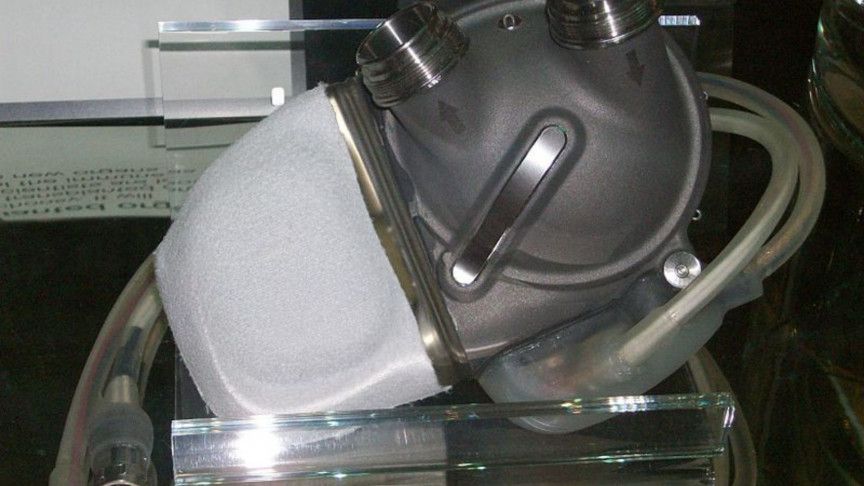
For more than 50 years, cardiac surgeons and biomedical engineers at the Texas Heart Institute (THI) have been questing for an artificial heart that can fully replace natural ones, which are in terribly short supply for transplant. They’ve seen their share of metal and plastic contraptions that used a variety of pumping mechanisms, but none of these machines could match the astounding performance of the human heart.

In April 2019, the possible culmination of that long quest was inside a shaggy brown cow, which stood peacefully chewing its cud at a THI research facility in Houston. The animal was part of a 90-day trial in which it lived its life powered by an implanted artificial heart made by our company, Bivacor. Throughout the trial, the calf stayed healthy and energetic, and gained weight at a normal rate. It even jogged on a treadmill for 30-minute stretches.

Our company is now working toward human trials of our device. It relies on a dramatic new approach: Rather than using a mechanical pump that mimics the structure and actions of the four-chambered human heart, it uses a spinning disk, suspended in a magnetic field. With just one moving part, the Bivacor heart is able to send oxygen-rich blood out to the body and return oxygen-depleted blood to the lungs.

Circa 2019


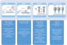
The evolution of micro and nanofabrication approaches significantly spurred the advancements of cardiac tissue engineering over the last decades. Engineering in the micro and nanoscale allows for the rebuilding of heart tissues using cardiomyocytes. The breakthrough of human induced pluripotent stem cells expanded this field rendering the development of human tissues from adult cells possible, thus avoiding the ethical issues of the usage of embryonic stem cells but also creating patient-specific human engineered tissues. In the case of the heart, the combination of cardiomyocytes derived from human induced pluripotent stem cells and micro/nano engineering devices gave rise to new therapeutic approaches of cardiac diseases. In this review, we survey the micro and nanofabrication methods used for cardiac tissue engineering, ranging from clean room-based patterning (such as photolithography and plasma etching) to electrospinning and additive manufacturing. Subsequently, we report on the main approaches of microfluidics for cardiac culture systems, the so-called “Heart on a Chip”, and we assess their efficacy for future development of cardiac disease modeling and drug screening platforms.

Circa 2020


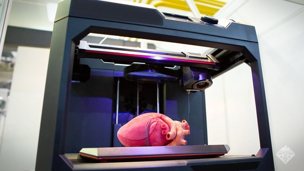
The FRESH technique of 3D bioprinting was invented in Feinberg’s lab to fill an unfilled demand for 3D printed soft polymers, which lack the rigidity to stand unsupported as in a normal print. FRESH 3D printing uses a needle to inject bioink into a bath of soft hydrogel, which supports the object as it prints. Once finished, a simple application of heat causes the hydrogel to melt away, leaving only the 3D bioprinted object.

While Feinberg, a professor of biomedical engineering and materials science and engineering, has proven both the versatility and the fidelity of the FRESH technique, the major obstacle to achieving this milestone was printing a human heart at full scale. This necessitated the building of a new 3D printer custom made to hold a gel support bath large enough to print at the desired size, as well as minor software changes to maintain the speed and fidelity of the print.

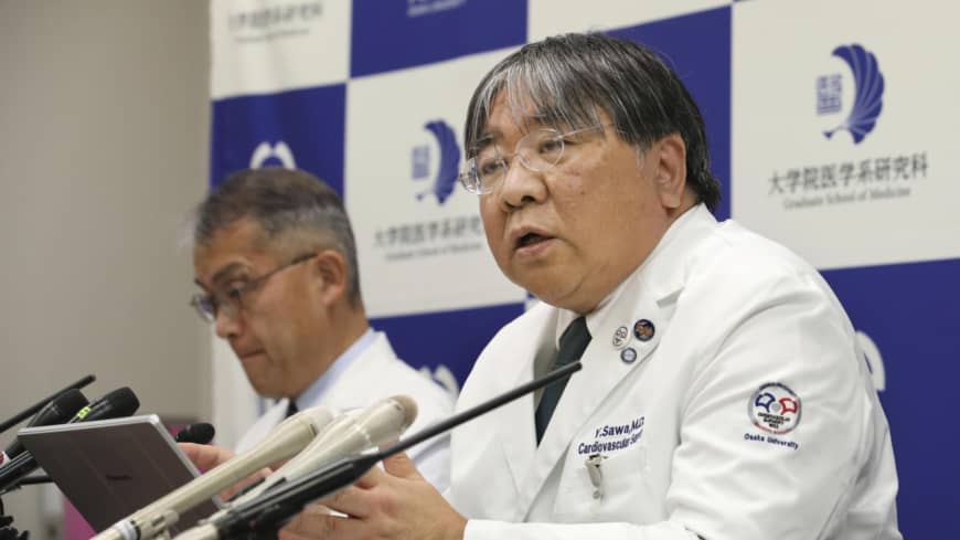
OSAKA – An Osaka University team said it has carried out the world’s first transplant of cardiac muscle cells created from iPS cells in a physician-initiated clinical trial.

In the clinical project to verify the safety and efficacy of the therapy using induced pluripotent stem cells, Yoshiki Sawa, a professor in the university’s cardiovascular surgery unit, and colleagues aim to transplant heart muscle cell sheets over the course of three years into 10 patients suffering from serious heart malfunction caused by ischemic cardiomyopathy.

Summary: Neuroinflammation may be a key player in the pathological brain changes produced as a result of chronic opioid use. Microglia is likely responsible for the majority of the changes.

Source: boston university school of medicine.

Prevalence rates of opioid use disorder (OUD) have increased dramatically, accompanied by a surge of overdose deaths–nearly 50000 in the U.S. in 2019. While opioid dependence has been extensively studied in preclinical models, an understanding of the biological alterations that occur in the brains of people who chronically use opioids and who are diagnosed with OUD remains limited.

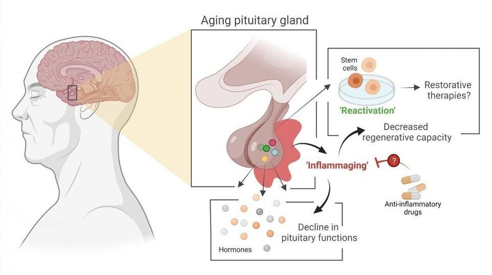
Stem cell biologist Hugo Vankelecom (KU Leuven) and his colleagues have discovered that the pituitary gland in mice ages as the result of an age-related form of chronic inflammation. It may be possible to slow down this process or even partially repair it. The researchers have published their findings in PNAS.

The pituitary is a small, globular gland located underneath the brain that plays a major role in the , explains Professor Hugo Vankelecom from the Department of Development and Regeneration at KU Leuven. “My research group discovered that the pituitary gland ages as a result of a form of chronic inflammation that affects tissue and even the organism as a whole. This usually goes unnoticed and is referred to as ‘inflammaging’—a contraction of inflammation and aging. Inflammaging has previously been linked to the aging of other organs.” Due to the central role played by the pituitary, its aging may contribute to the reduction of hormonal processes and hormone levels in our body—as is the case with menopause, for instance.

The study also provides significant insight into the stem cells in the aging . In 2012, Vankelecom and his colleagues showed that a prompt reaction of these stem cells to injury in the gland leads to repair of the tissue, even in adult animals. “As a result of this new study, we now know that stem cells in the pituitary do not lose this regenerative capacity when the organism ages. In fact, the stem cells are only unable to do their job because, over time, the pituitary becomes an ‘inflammatory environment’ as a result of the chronic inflammation. But as soon as the stem cells are taken out of this environment, they show the same properties as stem cells from a young pituitary.”