‘The most radical new university to open in decades.’ — The Times. Teaching students to tackle the world’s most complex problems.

Dr. James Giordano, Chief of the Neuroethics Studies Program and Scholar-in-Residence in the Pellegrino Center for Clinical Bioethics at Georgetown University, speaks to cadets and faculty about how advancements in neuroscience and neurotechnology will impact the future of war. This event was hosted by the Modern War Institute at West Point.
It’s not random.
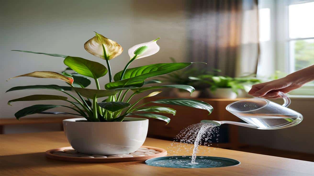
The Spathiphyllum is forgiving, but it signals stress fast. Brown patches and crunchy tips are a nudge from the plant about its air, water, or light. Here’s how specialists diagnose those signals and the small adjustments that bring foliage back to lush green.
Brown on a peace lily is usually a care mismatch, not a fatal disease. The plant is reacting to dry air, irregular watering, hard tap water, or harsh sun. Each cause marks leaves a little differently, which helps you zero in on the fix.
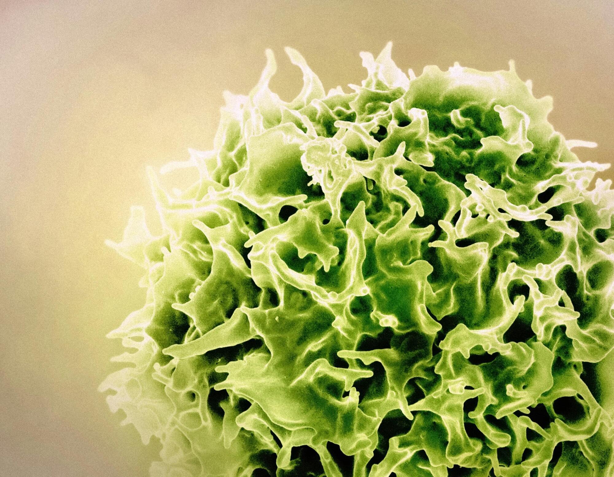
We owe a lot to tissue resident memory T cells (TRM). These specialized immune cells are among the body’s first responders to disease.
Rather than coursing through the bloodstream—as many T cells do—our TRM cells specialize in defending specific organs. They battle viruses, breast cancer, liver cancer, melanomas, and many other health threats.
Pandurangan Vijayanand, M.D., Ph.D., William K. Bowes Distinguished Professor at La Jolla Institute for Immunology (LJI), has even shown that a greater density of TRM cells is linked to better survival outcomes in lung cancer patients.
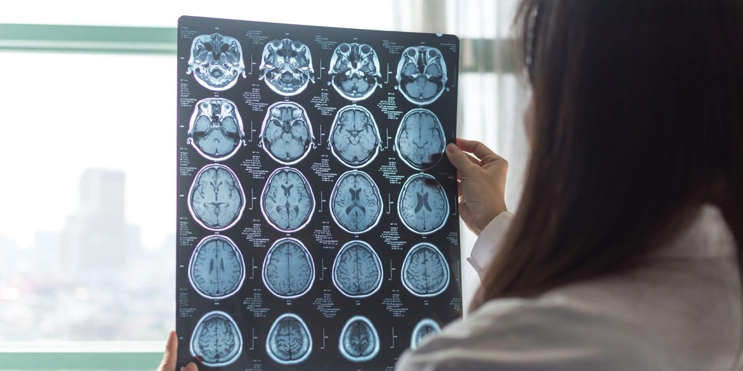
The study also utilized blood samples collected at the beginning of the research period. The investigators measured several biomarkers of systemic inflammation, including C-reactive protein and white blood cell counts. These markers were combined into a composite inflammation score. This score allowed the team to test if inflammation in the body explained the link between diet and brain changes.
The results revealed a significant association between dietary habits and brain aging. Individuals who consumed the most pro-inflammatory diets had a larger brain age gap compared to those who ate anti-inflammatory diets. Specifically, those in the most pro-inflammatory group had brains that appeared about half a year older on average than those in the healthiest diet group. This suggests that poor dietary quality may accelerate the biological clock of the brain.
This association was dependent on the age of the participants. The link between a pro-inflammatory diet and older brain age was much stronger in adults aged 60 and older. In this older demographic, a pro-inflammatory diet was associated with an advanced brain age of nearly a full year. This implies that older adults might be more vulnerable to the negative effects of a poor diet.
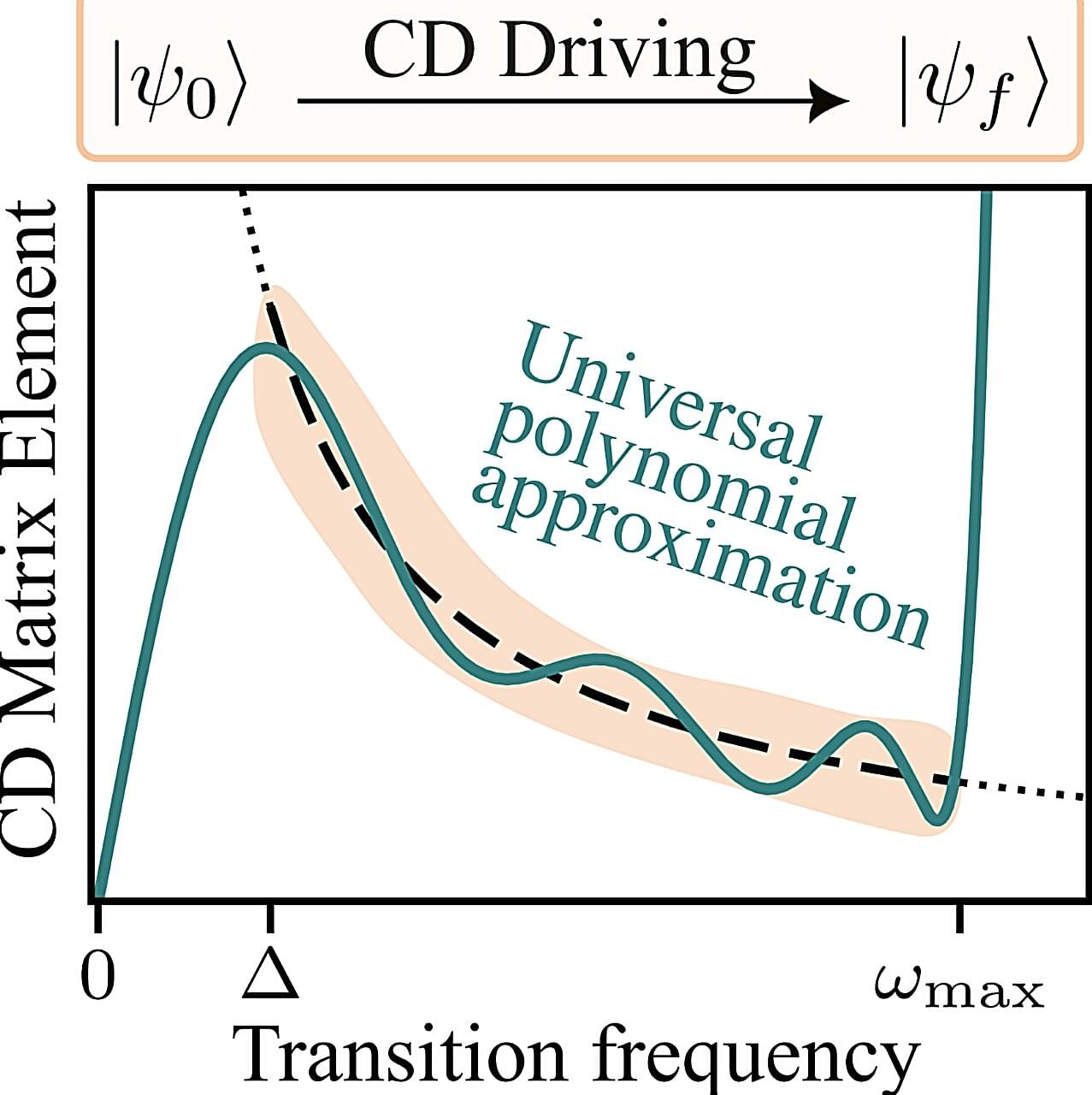
Quantum ground states are the states at which quantum systems have the minimum possible energy. Quantum computers are increasingly being used to analyze the ground states of interesting systems, which could in turn inform the design of new materials, chemical compounds, pharmaceutical drugs and other valuable goods.
The reliable preparation of quantum ground states has been a long-standing goal within the physics research community. One quantum computing method to prepare ground states and other desired states is known as adiabatic state preparation.
This is a process that starts from an initial Hamiltonian, a mathematical operator that encodes a system’s total energy and for which the ground state is known, gradually changing it to reach a final Hamiltonian, which encodes the final ground state.
Join us on Patreon! https://www.patreon.com/MichaelLustgartenPhD
Discount Links/Affiliates:
Blood testing (where I get the majority of my labs): https://www.ultalabtests.com/partners/michaellustgarten.
At-Home Metabolomics: https://www.iollo.com?ref=michael-lustgarten.
Use Code: CONQUERAGING At Checkout.
Clearly Filtered Water Filter: https://get.aspr.app/SHoPY
Epigenetic, Telomere Testing: https://trudiagnostic.com/?irclickid=U-s3Ii2r7xyIU-LSYLyQdQ6…M0&irgwc=1
Use Code: CONQUERAGING
NAD+ Quantification: https://www.jinfiniti.com/intracellular-nad-test/
Physicists have created a visible, self-sustaining “time crystal” using swirling liquid crystals that move in endlessly repeating patterns when illuminated. Imagine a clock that runs forever without batteries or wiring, its hands turning on their own without stopping. In a recent study, physic
Researchers discovered that the common amino acid arginine can block harmful Aβ aggregation and reduce its toxic effects in Alzheimer’s disease models. In flies and mice, oral arginine lowered plaque levels, reduced inflammation, and improved behavior. Its strong safety record and low cost make it a promising repurposing candidate. The findings hint at a surprisingly simple path toward more accessible AD therapies.
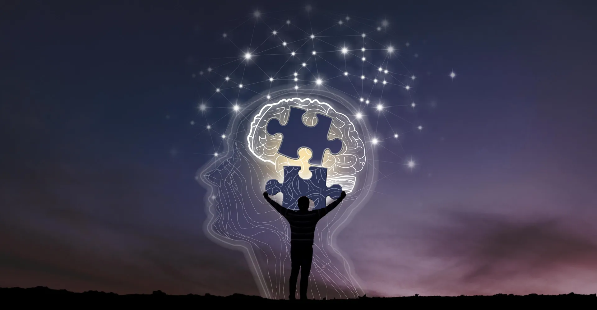
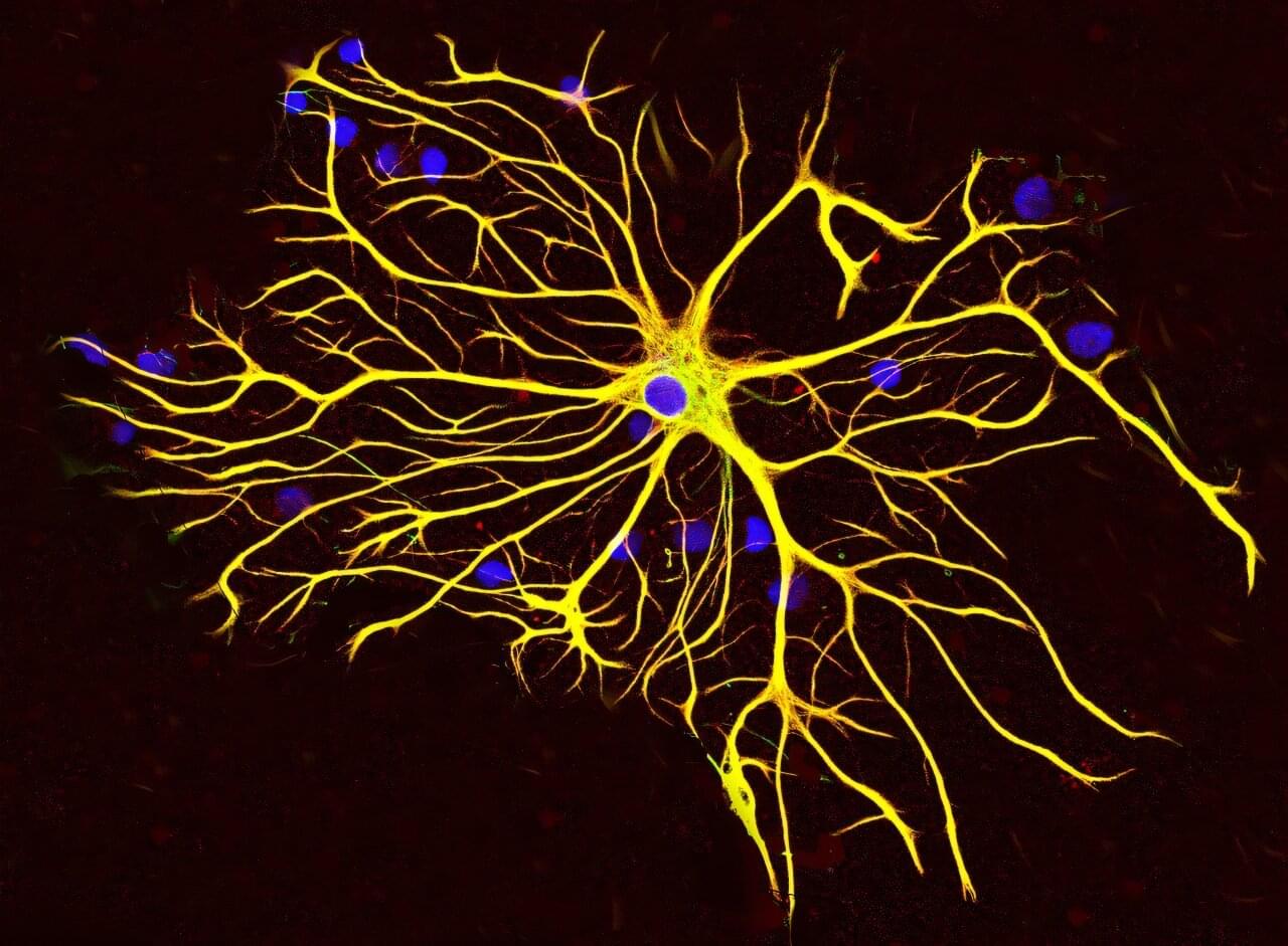
Researchers at Baylor College of Medicine have discovered a natural mechanism that clears existing amyloid plaques in the brains of mouse models of Alzheimer’s disease and preserves cognitive function. The mechanism involves recruiting brain cells known as astrocytes, star-shaped cells in the brain, to remove the toxic amyloid plaques that build up in many Alzheimer’s disease brains.
Increasing the production of Sox9, a key protein that regulates astrocyte functions during aging, triggered the astrocytes’ ability to remove amyloid plaques. The study, published in Nature Neuroscience, suggests a potential astrocyte-based therapeutic approach to ameliorate cognitive decline in neurodegenerative disease.
“Astrocytes perform diverse tasks that are essential for normal brain function, including facilitating brain communications and memory storage. As the brain ages, astrocytes show profound functional alterations; however, the role these alterations play in aging and neurodegeneration is not yet understood,” said first author Dr. Dong-Joo Choi, who was at the Center for Cell and Gene Therapy and the Department of Neurosurgery at Baylor while he was working on this project. Choi currently is an assistant professor at the Center for Neuroimmunology and Glial Biology, Institute of Molecular Medicine at the University of Texas Health Science Center at Houston.