The wildlife overpass spans Interstate 25 and connects 39,000 acres of habitat, allowing elk, deer, bears, mountain lions and other animals to cross safely.
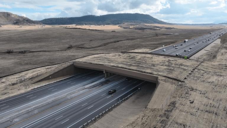
Sadly quite a realistic view.
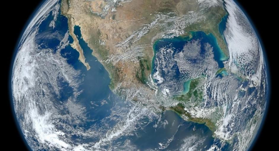
How special are we? A recent research paper suggests that terrestrial-style biology may be rare, and Earth may be among the first examples of a planet able to sustain life in the cosmos. Even as the new kids on the block, humans are seemingly one of the precious few instances of intelligence to arise in the universe since the Big Bang did its thing.
Related: The Search For Life Starts With Human Missions to Mars
Harvard Astronomer Avi Loeb and his colleagues in the U.K. have argued that the halcyon days for life are still to come. It’s not even morning in the universe; it’s pre-dawn. Biology may erupt like weeds on an untold number of worlds, but if so, the infestation will take place tens of billions of years in the future.
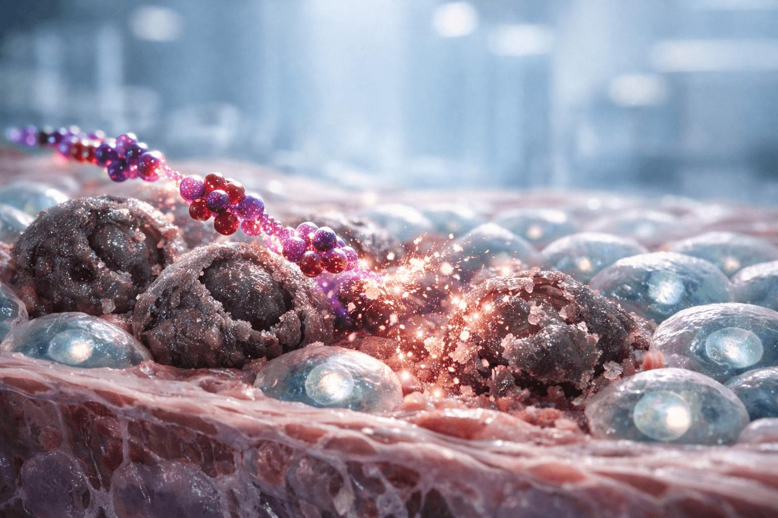
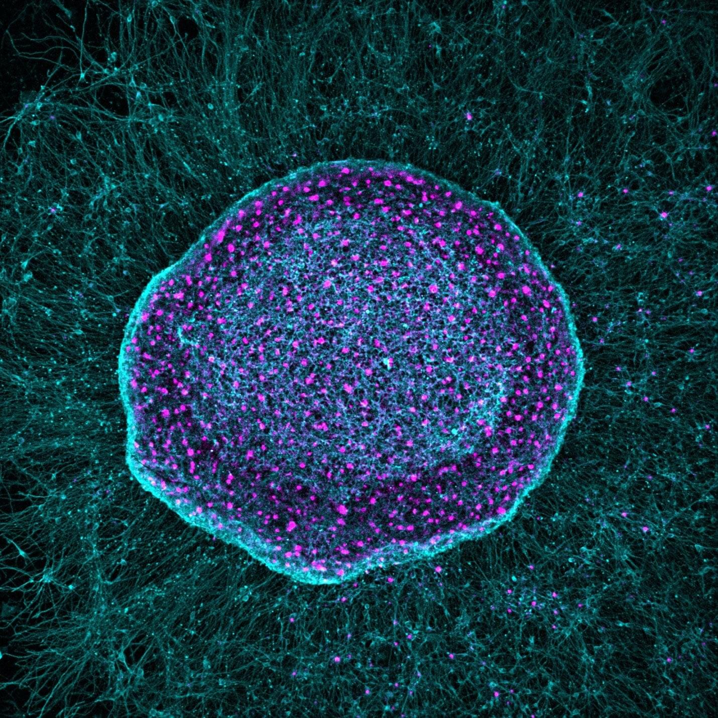
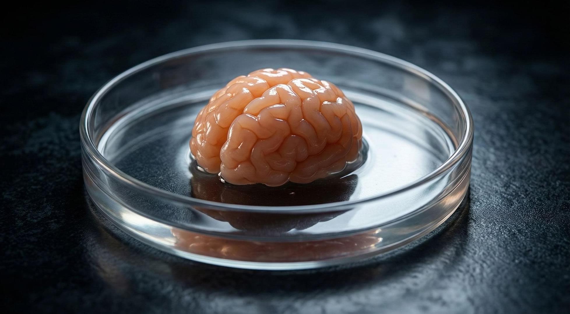
An Australian startup has unveiled the world’s first commercial biological computer (CL1), running on living human cells. CL1 is claimed to be capable of learning and adapting faster than standard silicon-based AI.
#News #ai #technology #futureofhealth #computer #braincells #Reuters #Newsfeed.
Read the story here:
👉 Subscribe: http://smarturl.it/reuterssubscribe.
Keep up with the latest news from around the world: https://www.reuters.com/
Follow Reuters on Facebook: / reuters.
Follow Reuters on Twitter: / reuters.
Follow Reuters on Instagram: https://www.instagram.com/reuters/?hl=en
Ozone depletion, economic crash, loss of access to space, these and many other threats from space debris can averted. See how turn space trash into cold cash!
GoldBacks from Galactic/Green Greg’s affiliate link:
https://www.defythegrid.com/goldbacks… coupon code GreenGregs for 1% off.

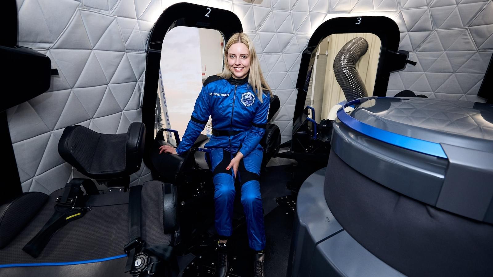
Blue Origin will make history when it sends the first person who uses a wheelchair past the Kármán line, an internationally recognized boundary of space that’s 62 miles above Earth.
On its next mission, a Blue Origin New Shepard rocket will take Michaela “Michi” Benthaus, an aerospace and mechatronics engineer who suffered a spinal cord injury after a mountain biking accident, along with five others, on a journey past the Kármán line. New Shepard rockets are fully reusable spacecraft that Blue Origin says require less maintenance between flights, saving money and reducing waste.
The NS-37 mission will be the 16th human flight for Blue Origin, which has taken 86 people — 80 individuals — above the Kármán line.