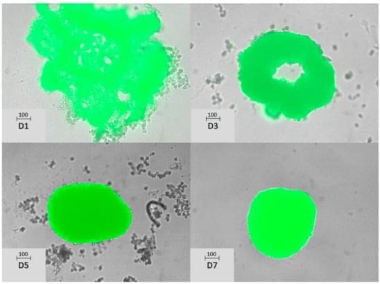
Background: Three-dimensional cellular models provide a more comprehensive representation of in vivo cell properties, encompassing physiological characteristics and drug susceptibility. Methods: Primary hepatocytes were seeded in ultra-low attachment plates to form spheroids, with or without tumoral cells. Spheroid structure, cell proliferation, and apoptosis were analyzed using histological staining techniques. In addition, extracellular vesicles were isolated from conditioned media by differential ultracentrifugation. Spheroids were exposed to cytotoxic drugs, and both spheroid growth and cell death were measured by microscopic imaging and flow cytometry with vital staining, respectively. Results: Concerning spheroid structure, an active outer layer forms a boundary with the media, while the inner core comprises a mass of cell debris.
