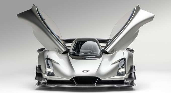
Toggle light / dark theme

Scientists build subcellular map of entire brain networks

Researchers at the Francis Crick Institute have developed an imaging technique to capture information about the structure and function of brain tissue at subcellular level—a few billionths of a meter, while also capturing information about the surrounding environment.

The unique approach detailed in Nature Communications today (25 May), overcomes the challenges of imaging tissues at different scales, allowing scientists to see the surrounding cells and how they function, so they can build a complete picture of neural networks in the .

Various imaging methods are used to capture information about , cells and subcellular structures. However, a single method can only capture information about either the structure or function of the tissue and looking in detail at a nanometer scale means scientists lose information about the wider surroundings. This means that to gain an overall understanding of the tissue, imaging techniques need to be combined.

Mapping functional connectivity in 3D artificial brain model

The human brain is less accessible than other organs because it is covered by a thick, hard skull. As a result, researchers have been limited to low-resolution imaging or analysis of brain signals measured outside the skull. This has proved to be a major hindrance in brain research, including research on developmental stages, causes of diseases, and their treatments. Recently, studies have been performed using primary neurons from rats or human-derived induced pluripotent stem cells (iPSCs) to create artificial brain models that have been applied to investigate brain developmental processes and the causes of brain diseases. These studies are expected to play a key role to unlocking the mysteries of the brain.

In the past, artificial models were created and studied in 2D; however, in 2017, a research team from KIST developed a 3D artificial brain model that more closely resembled the real brain. Unfortunately, due to the absence of an analytical framework for studying signals in a 3D brain model, studies were limited to analyses of surface signals or had to reform the 3D structure to a flat shape. As such, tracking in a complex, interconnected artificial network remained a challenge.

The Korea Institute of Science and Technology (KIST) announced that the research teams of Doctors Il-Joo Cho and Nakwon Choi have developed a that can apply precise non-destructive stimuli to a 3D artificial neural circuit and measure neural signals in real-time from multiple locations inside the model at the cellular level.

A hyperparameter optimization library for reproducible research

The table also shows the average normalized rank of transfer learning approaches. Hyperparameter transfer learning uses evaluation data from past HPO tasks in order to warmstart the current HPO task, which can result in significant speed-ups in practice.

Syne Tune supports transfer-learning-based HPO via an abstraction that maps a scheduler and transfer learning data to a warmstarted instance of the former. We consider the bounding-box and quantile-based ASHA, respectively referred to as ASHA-BB and ASHA-CTS. We also consider a zero-shot approach (ZS), which greedily selects hyperparameter configurations that complement previously considered ones, based on historical performances; and RUSH, which warmstarts ASHA with the best configurations found for previous tasks. As expected, we find that transfer learning approaches accelerate HPO.

Our experiments show that Syne Tune makes research on automated machine learning more efficient, reliable, and trustworthy. By making simulation on tabulated benchmarks a first-class citizen, it makes hyperparameter optimization accessible to researchers without massive computation budgets. By supporting advanced use cases, such as hyperparameter transfer learning, it allows better problem solving in practice.

Pinpointing Consciousness in Animal Brain Using Mouse ‘Brain Map’

Summary: Brain mapping study identifies important neural networks and their connections that appear to enhance the conscious experience.

Source: University of Tokyo

Science may be one step closer to understanding where consciousness resides in the brain. A new study shows the importance of certain types of neural connections in identifying consciousness.

The research, published in Cerebral Cortex, was led by Jun Kitazono, a corresponding author and a project researcher in the Department of General Systems Studies at the University of Tokyo.

NASA greenlights two new Mars helicopters and lengthens Perseverance’s resume

NASA and the European Space Agency (ESA) have agreed to “significant and advantageous changes” to a major part of the conceptual design for its Perseverance mission, NASA associate administrator Thomas Zurburchen states in the recent announcement.

This car-sized rover is the newest member of NASA’s robotic Mars fleet, and reached the Red Planet in February 2021 through an unprecedented landing. Arguably one of its most important responsibilities is the Mars Sample Return campaign. Perseverance’s six wheels leave grooves on the planet’s regolith as it works towards that goal, traversing Mars’ Jezero Crater to gather the telltale sedimentary proof that water — and possibly life — once existed there.

In October, the space agencies will dive into the details of their redesign: rather than having Perseverance leave caches of its pebble collection on Mars’ surface for another yet-to-be-built land-based spacecraft to pick up, the existing Mars rover will be the one to carry the precious parcels to their launch site. In addition, Perseverance’s high-flying robotic companion, the Ingenuity helicopter, has inspired the design of two future rotorcraft that would swerve over the Martian terrain to pick up other samples. This duo would be part of an existing concept, NASA’s Sample Retrieval Lander.

DeepMind’s AI has now catalogued every protein known to science

In late 2020, Alphabet’s DeepMind division unveiled its novel protein fold prediction algorithm, AlphaFold, and helped solve a scientific quandary that had stumped researchers for half a century. In the year since its beta release, half a million scientists from around the world have accessed the AI system’s results and cited them in their own studies more than 4,000 times. On Thursday, DeepMind announced that it is increasing that access even further by radically expanding its publicly-available AlphaFold Protein Structure Database (AlphaFoldDB) — from 1 million entries to 200 million entries.

Alphabet partnered with EMBL’s European Bioinformatics Institute (EMBL-EBI) for this undertaking, which covers proteins from across the kingdoms of life — animal, plant, fungi, bacteria and others. The results can be viewed on the UniProt, Ensembl, and OpenTargets websites or downloaded individually via GitHub, “for the human proteome and for the proteomes of 47 other key organisms important in research and global health,” per the AlphaFold website.

“AlphaFold is the singular and momentous advance in life science that demonstrates the power of AI,” Eric Topol, Founder and Director of the Scripps Research Translational Institute, siad in a press statement Thursday. “Determining the 3D structure of a protein used to take many months or years, it now takes seconds. AlphaFold has already accelerated and enabled massive discoveries, including cracking the structure of the nuclear pore complex. And with this new addition of structures illuminating nearly the entire protein universe, we can expect more biological mysteries to be solved each day.”

A “Nano-Robot” Built Entirely from DNA to Explore Cell Processes

Constructing a tiny robot from DNA and using it to study cell processes invisible to the naked eye… You would be forgiven for thinking it is science fiction, but it is in fact the subject of serious research by scientists from Inserm, CNRS and Université de Montpellier at the Structural Biology Center in Montpellier[1]. This highly innovative “nano-robot” should enable closer study of the mechanical forces applied at microscopic levels, which are crucial for many biological and pathological processes. It is described in a new study published in Nature Communications.

Our cells are subject to mechanical forces exerted on a microscopic scale, triggering biological signals essential to many cell processes involved in the normal functioning of our body or in the development of diseases.

For example, the feeling of touch is partly conditional on the application of mechanical forces on specific cell receptors (the discovery of which was this year rewarded by the Nobel Prize in Physiology or Medicine).