Toggle light / dark theme

Patient Number One is a thin man, with a scabby face and bouncy knees. His head, shaved in preparation for surgery, is wrapped in a clean, white cloth.

Years of drug use cost him his wife, his money and his self-respect, before landing him in this drab yellow room at a Shanghai hospital, facing the surgeon who in 72 hours will drill two small holes in his skull and feed electrodes deep into his brain.

The hope is that technology will extinguish his addiction, quite literally, with the flip of a switch.

Read more

Neurodegenerative diseases like multiple sclerosis (MS) affect millions of people worldwide and occur when parts of the nervous system lose function over time. Researchers at the University of Maryland School of Medicine (UMSOM) have discovered that a type of skin-related stem cell could be used to help regenerate myelin sheaths, a vital part of the nervous system linked to neurodegenerative disorders.

The discovery into these types of stem cells is significant because they could offer a simpler and less invasive alternative to using embryonic stem cells. This early stage research showed that by using these skin-related stem cells, researchers were able to restore myelin sheath formation in mice.

“This research enhances the possibility of identifying human skin stem cells that can be isolated, expanded, and used therapeutically. In the future, we plan to continue our research in this area by determining whether these cells can enhance functional recovery from neuronal injury,” said Thomas J. Hornyak, MD, PhD, Associate Professor and Chairman of the Department of Dermatology, and Principal Investigator in this research. “In the future, we plan to continue our research in this area by determining whether these cells can enhance functional recovery from neuronal injury.”

Read more

Brains contain a variety of immune cells that play an important role for brain function. A team led by Prof. Kiavash Movahedi (VIB Center for Inflammation Research at VUB) has developed a comprehensive cell atlas of the brain’s immune compartment. This revealed not only the striking diversity of brain macrophages, but also uncovered microglia where they were not expected. Remarkably, these previously unknown microglia showed a clear resemblance to microglia that are normally associated with neurodegenerative disorders such as Alzheimer’s disease. The new insights are important for understanding the role of macrophages in healthy brain physiology and for developing future treatments for neurodegenerative diseases.

Macrophages in the brain were first discovered 100 years ago by the Spanish scientist P\xEDo del R\xEDo-Hortega. Most brain macrophages are known as . These cells are in close contact with neurons and are critical for the proper development and functioning of the brain. But beyond the microglia, brains house several other types of macrophages, many of which are relatively unknown.

Prof. Kiavash Movahedi (VIB Center for Inflammation Research, VUB), said, “While microglia are fairly well studied, other brain macrophages have remained quite enigmatic. We wanted to obtain a better understanding of these cells, as we believe they could be critical for regulating brain inflammation and immunity.”

Read more

Scientists from Trinity College Dublin have discovered a potential new target for regulating inflammation, which drives a range of diseases including diabetes, cancer and Alzheimer’s. The potential target is an ancient immune protein—SARM—that has been conserved throughout evolution and thus is very similar in humans, other mammals, flies and worms.

The scientists, from Trinity’s School of Biochemistry and Immunology based at the Trinity Biomedical Sciences Institute (TBSI), discovered a previously unknown but important role that SARM plays in the . Their work has been published today in the prestigious journal Immunity.

Read more

A discovery opens the possibility of one day restoring loss of vision by activating the retina’s ability to regenerate. Researchers at Baylor College of Medicine, the Cardiovascular Research Institute and the Texas Heart Institute reveal in the journal Cell Reports that although the mammalian retina—a layer of specialized nerve cells that mediates vision and is located on the back of the eye- does not spontaneously regenerate, it has a regenerative capacity that is kept dormant by a cellular mechanism called the Hippo pathway. The discovery opens the possibility of activating the retina’s ability to restore lost vision by manipulating this pathway.

“Damage to the can lead to irreparable loss of vision in humans and other mammals because their retinas do not regenerate,” said lead author Dr. Ross A. Poché, assistant professor of molecular physiology and biophysics and member of the Dan L Duncan Comprehensive Cancer Center at Baylor. “However, other animals such as zebrafish can reverse blindness thanks to specialized cells in the retina called Müller . When the retina is damaged, Müller glial cells proliferate and differentiate into the lost , effectively replacing injured cells with fully functional ones.”

Although Müller glial cells in injured mammalian retina do not restore vision as their counterpart in zebrafish do, other researchers have shown that, when the mammalian retina is injured, a small subset of Müller glial cells takes the first steps needed to enter the proliferation cycle, such as acquiring molecular markers scientists expect to see in a proliferating cell.

Read more

SpiNNaker was built under the leadership of Professor Steve Furber at The University of Manchester, a principal designer of two products that earned the Queen’s Award for Technology —the ARM 32-bit RISC microprocessor, and the BBC Microcomputer.

“The ultimate objective for the project has always been a million cores in a single computer for real time brain modelling applications, and we have now achieved it, which is fantastic.” — Professor Steve Furber, The University of Manchester

Inspired by the human brain, the SpiNNaker is capable of sending billions of small amounts of information simultaneously. The SpiNNaker has a staggering 1 million processors that are able to perform over 200 million actions per second.

Read more

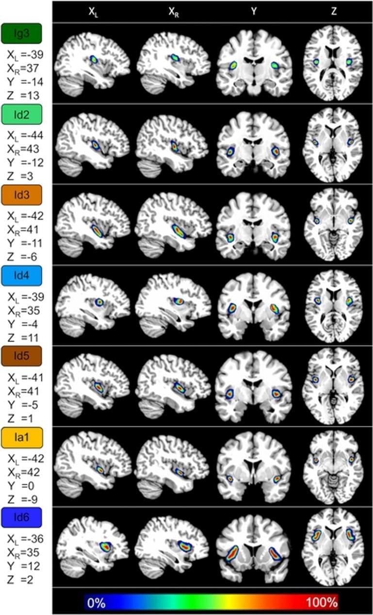
Neuroscience, computer vision collaborate to better understand visual information processing PITTSBURGH—Neuroscientists and computer vision scientists say a new dataset of unprecedented size — comprising brain scans of four volunteers who each viewed 5,000 images — will help researchers better understand how the brain processes images. Researchers at Carnegie Mellon University and Fordham University, reporting today in the journal Scientific Data, said acquiring functional magnetic resonance imaging (fMRI) scans at this scale presented unique challenges. Each volunteer participated in 20 or more hours of MRI scanning, challenging both their perseverance and the experimenters’ ability to coordinate across scanning sessions. The extreme.