In this exclusive excerpt from ‘Your Brain on Art,’ we learn how sounds and images are proving to measurably heal the brain.
Changes in the brain caused by Alzheimer’s disease are associated with shortening of the telomeres—the protective caps on the ends of chromosomes that shorten as cells age—according to a new study led by Anya Topiwala of Oxford Population Health, part of the University of Oxford, UK, published March 22 in the open-access journal PLOS ONE.
Telomeres on chromosomes protect DNA from degrading, but every time a cell divides, the telomeres lose some of their length. Short telomeres are a sign of stress and cellular aging, and are also associated with a higher risk of neurological and psychiatric disorders. Currently, little is known about the links between telomere length and changes that occur in the brains of people with neurological conditions. Understanding those relationships could offer insights into the biological mechanisms that cause neurodegenerative disorders.
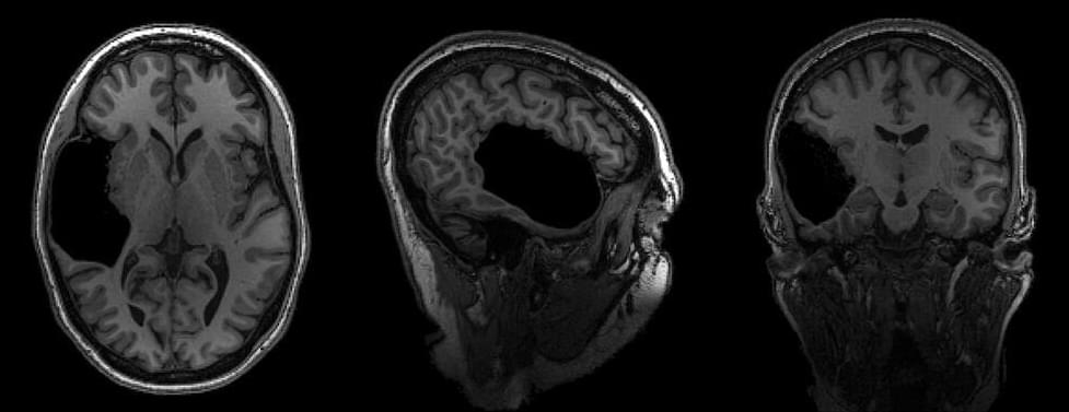
In the new study, researchers compared telomere length in white blood cells to results from brain MRIs and electronic health records from more than 31,000 participants in the UK Biobank, a large-scale biomedical database and research resource containing anonymized genetic, lifestyle and health information from half a million UK participants.
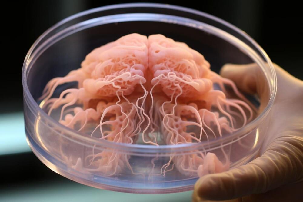
Watch as primordial neural cells dance across, grow into, and even move 3D scaffolds engineered to heal brain injury from stroke and other trauma. Decorating the scaffold with various nutrients and biochemical signals allow researchers to control what types of brain tissues they become.
“This interface could revolutionize the way we interact with technology.”
Researchers from the University of Cambridge have created a new type of neural implant that could restore limb function in paralyzed limbs.
There have been former attempts at using neural implants to restore limb function, but these mostly failed. This is because scar tissue can envelop the electrodes over time, disrupting the connection between the device and the nerve.
University of Cambridge.
The developed device works in sync between the brain and paralyzed limbs — it combines flexible electronics and human stem cells to “better integrate” with the nerve and drive limb function, according to a press release.
Near Death Experience Research Foundation the largest collection of Near Death Experiences (NDE) in over 23 Languages. With thousands of full-text near death experiences posted. Share your near death experience, research, spiritually transforming events, consciousness studies, extensive information and research.
In this video, we will explore the positional system of the brain — hippocampal place cells. We will see how it relates to contextual memory and mapping of more abstract features.
OUTLINE:
00:00 Introduction.
00:53 Hippocampus.
1:27 Discovery of place cells.
2:56 3D navigation.
3:51 Role of place cells.
4:11 Virtual reality experiment.
7:47 Remapping.
11:17 Mapping of non-spatial dimension.
13:36 Conclusion.
_____________
REFERENCES:
1) Anderson, M.I., Jeffery, K.J., 2003. Heterogeneous Modulation of Place Cell Firing by Changes in Context. J. Neurosci. 23, 8827–8835. https://doi.org/10.1523/JNEUROSCI.23-26-08827.
2) Aronov, D., Nevers, R., Tank, D.W., 2017. Mapping of a non-spatial dimension by the hippocampal–entorhinal circuit. Nature 543719–722. https://doi.org/10.1038/nature21692
3) Bostock, E., Muller, R.U., Kubie, J.L., 1991. Experience-dependent modifications of hippocampal place cell firing. Hippocampus 1193-205. https://doi.org/10.1002/hipo.
For individuals suffering from drug addiction, certain cues—whether it’s specific people, places or things—can trigger powerful cravings for repeated use.
A new University of Michigan study has identified brain signals, traditionally associated with inflammation, contributing to people’s vulnerability to addiction. With repeated drug use with the same exposure to cues, some individuals develop an inability to control their drug use, even in the face of negative consequences.
The study is published in the journal eNeuro.
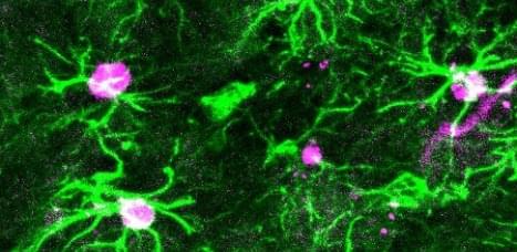
The study authors claim their microscope can provide colored real-time images of hard-to-reach parts of the spinal cord that couldn’t be accessed previously.
Pain is a powerful feeling but have you ever wondered how pain works on a cellular level? Well, a team of scientists at the San Diego-based Salk Insitute has actually figured out a way to see the internal neural mechanism associated with pain.
In their recently published study, they propose wearable microscopes using which they were able to check how nerve cells in the spinal cord of mice process pain signals.
Salk Institute.