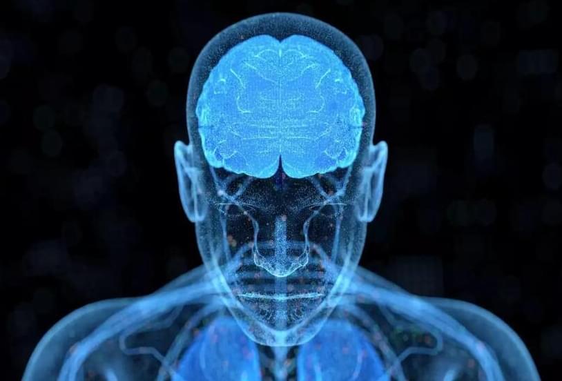
Magnetic stimulation is helping some people with depression—but the $12,000 treatment is also being unleashed in untested ways.

Circa 2013
“When the induced field is above a certain threshold, and is directed in an appropriate orientation relative to the brain’s neuronal pathways, localized axonal depolarizations are produced, thus activating the neurons in the relevant brain structure.”
First the machine is calibrated by placing it over a part of the brain that causes the subject’s hand to move. Then the coils are aimed at the brain region under treatment. The treatment lasts about 15 to 30 minutes, repeated over several weeks, and is noninvasive–all the person feels is a slight buzzing, and there are no side effects. This makes it a more palatable relative of other treatments that also target the brain directly, such as electroconvulsive therapy (formerly electroshock), or surgically implanted electrodes.
Summary: Boosting levels of the DUSP4 protein could be a novel way of preventing and treating epilepsy.
Source: University of Illinois
Epileptic seizures often originate in small, localized areas of the brain where neurons abnormally fire in unison. These electrical impulses disrupt proper brain functioning and cause seizures. But what makes regions where seizures start different from parts of the brain where electrical impulses remain normal? More importantly, what prevents these epileptic centers from growing?
Researchers have made a breakthrough genetic discovery into the cause of a spectrum of severe neurological conditions.
A research study, led by the Murdoch Children’s Research Institute (MCRI) and gracing the cover of and published in the October edition of Human Mutation, found two new mutations in the KIF1A gene cause rare nerve disorders.
MCRI researcher Dr. Simranpreet Kaur said mutations in the KIF1A gene caused ‘traffic jams’ in brain cells, called neurons, triggering a devastating range of progressive brain disorders. KIF1A-Associated Neurological Disorders (KAND) affects about 300 children worldwide.
Imagine if your manager could know whether you actually paid attention in your last Zoom meeting. Or, imagine if you could prepare your next presentation using only your thoughts. These scenarios might soon become a reality thanks to the development of brain-computer interfaces (BCIs).
To put it in the simplest terms, think of a BCI as a bridge between your brain and an external device. As of today, we mostly rely on electroencephalography (EEG) — a collection of methods for monitoring the electrical activity of the brain — to do this. But, that’s changing. By leveraging multiple sensors and complex algorithms, it’s now becoming possible to analyze brain signals and extract relevant brain patterns. Brain activity can then be recorded by a non-invasive device — no surgical intervention needed. In fact, the majority of existing and mainstream BCIs are non-invasive, such as wearable headbands and earbuds.
The development of BCI technology was initially focused on helping paralyzed people control assistive devices using their thoughts. But new use cases are being identified all the time. For example, BCIs can now be used as a neurofeedback training tool to improve cognitive performance. I expect to see a growing number of professionals leveraging BCI tools to improve their performance at work. For example, your BCI could detect that your attention level is too low compared with the importance of a given meeting or task and trigger an alert. It could also adapt the lighting of your office based on how stressed you are, or prevent you from using your company car if drowsiness is detected.
One of the most remarkable recent advances in biomedical research has been the development of highly targeted gene-editing methods such as CRISPR that can add, remove, or change a gene within a cell with great precision. The method is already being tested or used for the treatment of patients with sickle cell anemia and cancers such as multiple myeloma and liposarcoma, and today, its creators Emmanuelle Charpentier and Jennifer Doudna received the Nobel Prize in chemistry.
While gene editing is remarkably precise in finding and altering genes, there is still no way to target treatment to specific locations in the body. The treatments tested so far involve removing blood stem cells or immune system T cells from the body to modify them, and then infusing them back into a patient to repopulate the bloodstream or reconstitute an immune response—an expensive and time-consuming process.
Building on the accomplishments of Charpentier and Doudna, Tufts researchers have for the first time devised a way to directly deliver gene-editing packages efficiently across the blood brain barrier and into specific regions of the brain, into immune system cells, or to specific tissues and organs in mouse models. These applications could open up an entirely new line of strategy in the treatment of neurological conditions, as well as cancer, infectious disease, and autoimmune diseases.
A team of researchers from University Hospital Zurich and the University of Zurich, the Swiss Federal Institute of Technology and biotechnology company Philochem, has found fusing cytokines with antibodies to be an effective treatment for glioblastoma in mice. In their paper published in the journal Science Translational Medicine, the group describes their technique and how well it worked when tested with mouse models.
Glioblastoma is a type of cancerous brain tumor that is notoriously difficult to treat. Surgery to remove it is difficult and fraught with side effects, and drugs have little impact. In recent years, researchers have tried using drugs that alert the immune system to the presence of the tumor but they have not worked as hoped, either. In this new effort, the researchers tried another approach—fusing cytokines with antibodies as a way to attack the tumor. The hope was that together, the two would incite the immune system to attack the tumor more strongly and hopefully get rid of it.
Cytokines are small protein cells secreted by the immune system. Their usual job is to send signals to other cells in the immune system. And antibodies are Y-shaped proteins produced by plasma cells, the workhorses of the immune system that attack viruses and bacteria. In this new effort, the researchers fused L19 antibodies to cytokines. L19 was chosen because prior research has shown that it is able to seek out markers for glioblastoma. Fusing the two proteins together proved to be a more formidable therapeutic approach than using either alone. The resulting (L19TNF) immunocytokines were injected into mice with induced glioblastoma and were then monitored to determine their impact on the brain tumors.