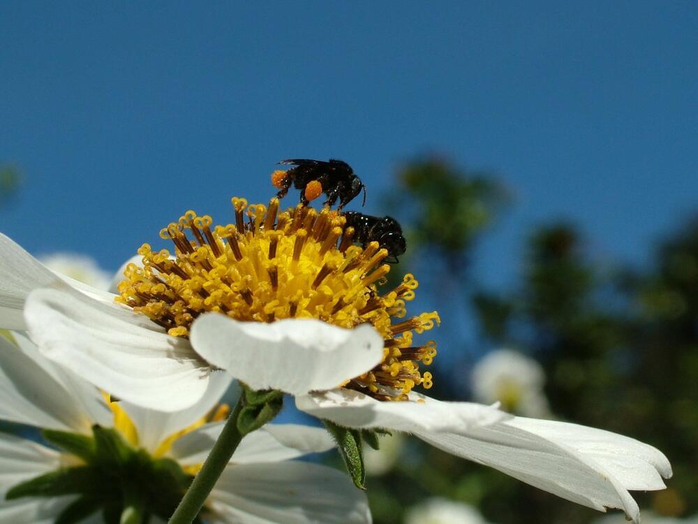
Toggle light / dark theme

A Duke University research team has found a small area of the brain in mice that can profoundly control the animals’ sense of pain.

Somewhat unexpectedly, this center turns off, not on. It’s also located in an area where few people would have thought to look for an anti-pain center, the amygdala, which is often considered the home of negative emotions and responses, like the fight or flight response and general anxiety.

“People do believe there is a central place to relieve pain, that’s why placebos work,” said senior author Fan Wang, the Morris N. Broad Distinguished Professor of neurobiology in the School of Medicine. “The question is where in the brain is the center that can turn off pain.”

A Duke University research team has found a small area of the brain in mice that can profoundly control the animals’ sense of pain.

Somewhat unexpectedly, this brain center turns pain off, not on. It’s also located in an area where few people would have thought to look for an anti-pain center, the amygdala, which is often considered the home of negative emotions and responses, like the fight or flight response and general anxiety.

“People do believe there is a central place to relieve pain, that’s why placebos work,” said senior author Fan Wang, the Morris N. Broad Distinguished Professor of neurobiology in the School of Medicine. “The question is where in the brain is the center that can turn off pain.”

Summary: New neurostimulation technology works safely and non-invasively to modify brain activity. The findings may provide some foundational knowledge for the development of future technologies that could expedite cognitive processes.

Source: US Army Research Laboratory

For Army scientists, the goal of neuroscience research is pursuing the inner workings of the human brain to advance scientific understanding and improve Soldier performance.

Monogamous prairie voles form lifelong pair bonds, but the neuronal dynamics that underlie bond formation and maintenance in this species remain largely unknown. We performed imaging of populations of neurons while voles interacted with their pair-bonded partner or a novel vole before and after bond formation. We identified neurons that were active during partner approach and found that this subset of cells was distinct from those that were active during novel approach. Furthermore, the number of partner approach cells increased following bond formation, reflecting the emergence of bonding behaviors and correlating with bond strength. This discovery sheds light on how pair bonds may be encoded within the brain and what changes as bonds mature.

Pair-bond formation depends vitally on neuromodulatory signaling within the nucleus accumbens, but the neuronal dynamics underlying this behavior remain unclear. Using 1-photon in vivo Ca2+ imaging in monogamous prairie voles, we found that pair bonding does not elicit differences in overall nucleus accumbens Ca2+ activity. Instead, we identified distinct ensembles of neurons in this region that are recruited during approach to either a partner or a novel vole. The partner-approach neuronal ensemble increased in size following bond formation, and differences in the size of approach ensembles for partner and novel voles predict bond strength. In contrast, neurons comprising departure ensembles do not change over time and are not correlated with bond strength, indicating that ensemble plasticity is specific to partner approach.

During sleep and awake rest, the neocortex generates large-scale slow-wave (SW) activity. Here, we report that the claustrum coordinates neocortical SW generation. We established a transgenic mouse line that enabled the genetic interrogation of a subpopulation of claustral glutamatergic neurons. These neurons received inputs from and sent outputs to widespread neocortical areas. The claustral neuronal firings mostly correlated with cortical SW activity. In vitro optogenetic stimulation of the claustrum induced excitatory postsynaptic responses in most neocortical neurons, but elicited action potentials primarily in inhibitory interneurons. In vivo optogenetic stimulation induced a synchronized down-state featuring prolonged silencing of neural activity in all layers of many cortical areas, followed by a down-to-up state transition. In contrast, genetic ablation of claustral neurons attenuated SW activity in the frontal cortex. These results demonstrate a crucial role of claustral neurons in synchronizing inhibitory interneurons across wide cortical areas for the spatiotemporal coordination of SW activity.

Scientists have discovered a new treatment to dramatically reduce swelling after brain and spinal cord injuries, offering hope to 75 million victims worldwide each year.

The breakthrough in treating such injuries—referred to as central nervous system (CNS) edema—is thought to be hugely significant because current options are limited to putting patients in an induced coma or performing risky surgery.

Brain and affect all age groups. Older people are more at risk of sustaining them from strokes or falls, while for younger age groups, major causes include road traffic accidents and injuries from sports such as rugby, US-style football and other contact games.

For Army scientists, the goal of neuroscience research is pursuing the inner workings of the human brain to advance scientific understanding and improve Soldier performance.

Researchers recently applied new techniques to modify brain activity. Not only are these techniques used to characterize and study complex networks such as in telecommunications or social networks—they describe how different nodes, or elements of the network: brain regions in neuroscience, or individuals in social networks, interact with each other.

The U.S. Army Combat Capabilities Development Command’s Army Research Laboratory, in collaboration with academic partners, collaborated on a neurostimulation study, where they safely and non-invasively modified and then characterized the dynamics of the brain’s response to this modification. This research provides some of the foundational knowledge for future technologies that may one day expedite cognitive processes. The journal Network Neuroscience published the recent discoveries.