UW Medicine researchers have found that algorithms are as good as trained human evaluators at identifying red-flag language in text messages from people with serious mental illness. This opens a promising area of study that could help with psychiatry training and scarcity of care.
The findings were published in late September in the journal Psychiatric Services.
Text messages are increasingly part of mental health care and evaluation, but these remote psychiatric interventions can lack the emotional reference points that therapists use to navigate in-person conversations with patients.
The brain’s ability to adapt and rewire itself throughout life continues to surprise neuroscientists. Researchers have found a way to restore sight in adult mice with a form of congenital blindness, in spite of the rodents’ relative maturity.
The mice were modeling a rare human disorder of the eye’s retina, called leber congenital amaurosis (LCA), which often causes blindness or severe visual impairment at birth.
This inherited condition seems to be caused by a mutation in any one of dozens of genes associated with the retina and its light-sensing abilities.
Michael Levin is a developmental and synthetic biologist at Tufts University, where he is the Vannevar Bush Distinguished Professor of biology. He is a director of the Allen Discovery Center, director at the Tufts Center for Regenerative and Developmental Biology, and principal investigator at the Levin Lab.
0:00 intro. 1:38 bioelectricity and developmental biology. 7:56 memory and conditioning in GRNs. 11:50 is there a privileged cognitive substrate? 13:55 Godel type limits. 15:45 multi-scale competency architecture. 25:12 intelligence. 27:00 conceptual framework for cognition. 29:45 does cognition bottom out somewhere? 36:47 synthetic cognition. 39:23 sci-fi that captures this well. 45:16 consciousness, hard problem, and the consciousness of development. 51:09 where does the self come from? 54:06 how do different emergent levels interact. 56:50 top-down causality. 1:02:28 where do goals come from? 1:07:06 balancing conceptual and empirical work.
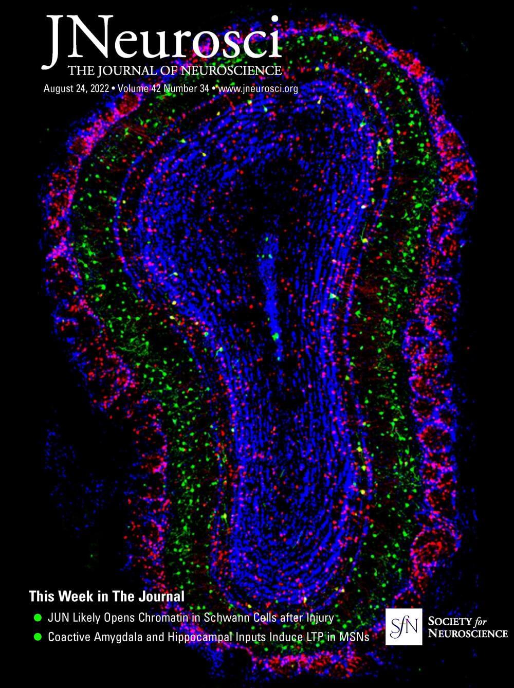
Large glutamatergic, somatic synapses mediate temporally precise information transfer. In the ventral nucleus of the lateral lemniscus, an auditory brainstem nucleus, the signal of an excitatory large somatic synapse is sign inverted to generate rapid feedforward inhibition with high temporal acuity at sound onsets, a mechanism involved in the suppression of spurious frequency information. The mechanisms of the synaptically driven input–output functions in the ventral nucleus of the lateral lemniscus are not fully resolved. Here, we show in Mongolian gerbils of both sexes that, for stimulation frequencies up to 200 Hz, the EPSC kinetics together with short-term plasticity allow for faithful transmission with only a small increase in latency. Glutamatergic currents are exclusively mediated by AMPARs and NMDARs. Short-term plasticity is frequency-dependent and composed of an initial facilitation followed by depression. Physiologically relevant output generation is limited by the decrease in synaptic conductance through short-term plasticity (STP). At this endbulb synapse, STP acts as a low pass filter and increases the dynamic range of the conductance dependent input–output relation, while NMDAR signaling slightly increases the sensitivity of the input–output function. Our computational model shows that STP-mediated filtering limits the intensity dependence of the spike output, thus maintaining selectivity to sound transients. Our results highlight the interaction of cellular features that together give rise to the computations in the circuit.
SIGNIFICANCE STATEMENT Auditory information processing in the brainstem is a prerequisite for generating our auditory representation of the environment. Thereby, many processing steps rely on temporally precise filtering. Precise feedforward inhibition is a key motif in auditory brainstem processing and produced through sign inversion at several large somatic excitatory synapses. A particular feature of the ventral nucleus of the lateral lemniscus is to produce temporally precise onset inhibition with little temporal variance independent of sound intensity. Our cell-physiology and modeling data explain how the synaptic characteristics of different current components and their short-term plasticity are tuned to establish sound intensity-invariant onset inhibition that is crucial for filtering out spurious frequency information.
Breakthrough AI programs can now generate videos from text input. The U.S. suicide rate and the prevalence of anxiety disorders are at all-time high. The White House has announced the “AI Bill of Rights.” What’s the connection between these 3 news items?
They all hint at how we will live our lives in the near future: As illusionists, making up imaginary worlds, fearing fabricated threats, led by conjurers, tricksters, and demagogues. For some, this prediction is already a good approximation of their present reality.
Let’s start with “AI,” the most exciting, confusing, and menacing technology of our times.
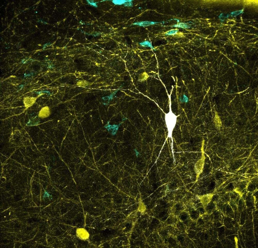
A study led by researchers from the Institute Cajal of Spanish Research Council (CSIC) in Madrid, Spain in collaboration with the Bioengineering Department of George Mason University in Virginia, U.S. has updated one of the world’s largest databases on neuronal types, Hippocampome.org.
The study, which is published in the journal PLOS Biology, represents the most comprehensive mapping performed to date between neural activity recoded in vivo and identified neuron types. This major breakthrough may enable biologically meaningful computer modeling of the full neuronal circuit of the hippocampus, a region of the brain involved in memory function.
Circuits of the mammalian cerebral cortex are made up of two types of neurons: Excitatory neurons, which release a neurotransmitter called glutamate, and inhibitory neurons, which release GABA (gamma-aminobutanoic acid), the main inhibitor of the central nervous system. “A balanced dialogue between the ‘excitatory’ and ‘inhibitory’ activities is critical for brain function. Identifying the contribution from the several types of excitatory and inhibitory cells is essential to better understand brain operation,” explains Liset Menendez de la Prida, the Director of the Laboratorio de Circuitos Neuronales at the Institute Cajal who leads the study at the CSIC.
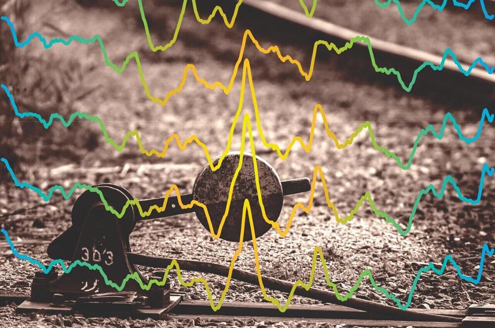
A team of scientists has uncovered a system in the brain used in the processing of information and in the storing of memories—akin to how railroad switches control a train’s destination. The findings offer new insights into how the brain functions.
“Researchers have sought to identify neural circuits that have specialized functions, but there are simply too many tasks the brain performs for each circuit to have its own purpose,” explains André Fenton, a professor of neural science at New York University and the senior author of the study, which appears in the journal Cell Reports. “Our results reveal how the same circuit takes on more than one function. The brain diverts ‘trains’ of neural activity from encoding our experiences to recalling them, showing that the same circuits have a role in both information processing and in memory.”
This newly discovered dynamic shows how the brain functions more efficiently than previously realized.
A paradigm shift in how we think about the functions of the human brain. A long-awaited genetic sequence of Rafflesia arnoldii, the strangest flower in the world. A revelation in sleep science. These are some of the year’s biggest discoveries in neuroscience and other areas of biology. Read the articles in full at Quanta: https://www.quantamagazine.org/the-year-in-biology-20211221/
Quanta Magazine is an editorially independent publication supported by the Simons Foundation.
In this episode we explore a User Interface Theory of reality. Since the invention of the computer virtual reality theories have been gaining in popularity, often to explain some difficulties around the hard problem of consciousness (See Episode #1 with Sue Blackmore to get a full analysis of the problem of how subjective experiences might emerge out of our brain neurology); but also to explain other non-local anomalies coming out of physics and psychology, like ‘quantum entanglement’ or ‘out of body experiences’. Do check the devoted episodes #4 and #28 respectively on those two phenomena for a full breakdown. As you will hear today the vast majority of cognitive scientists believe consciousness is an emergent phenomena from matter, and that virtual reality theories are science fiction or ‘Woowoo’ and new age. One of this podcasts jobs is to look at some of these Woowoo claims and separate the wheat from the chaff, so the open minded among us can find the threshold beyond which evidence based thinking, no matter how contrary to the consensus can be considered and separated from wishful thinking. So you can imagine my joy when a hugely respected cognitive scientist and User Interface theorist, who can cut through the polemic and orthodoxy with calm, respectful, evidence based argumentation, agreed to come on the show, the one and only Donald D Hoffman.
Hoffman is a full professor of cognitive science at the University of California, Irvine, where he studies consciousness, visual perception and evolutionary psychology using mathematical models and psychophysical experiments. His research subjects include facial attractiveness, the recognition of shape, the perception of motion and colour, the evolution of perception, and the mind-body problem. So he is perfectly placed to comment on how we interpret reality.
Hoffman has received a Distinguished Scientific Award of the American Psychological Association for early career research into visual perception, the Rustum Roy Award of the Chopra Foundation, and the Troland Research Award of the US National Academy of Sciences. So his recognition in the field is clear.
He is also the author of ‘The Case Against Reality’, the content of which we’ll be focusing on today; ‘Visual Intelligence’, and the co-author with Bruce Bennett and Chetan Prakash of ‘Observer Mechanics’.
What we discuss: 00:00 Intro. 05:30 Belief VS questioning. 11:20 Seeing the world for survival VS for knowing reality as it truly is. 13:30 Competing strategies to maximise ‘fitness’ in the evolutionary sense. 15:22 Fitness payoff’s can be calculated as mathematical functions, based on different organisms, states and actions. 17:00 Evolutionary Game Theory computer simulations at UC Irvine. 21:30 The payoff functions that govern evolution do not contain information about the structure of the world. 25:00 The world is NOT as it seems VS The world is NOTHING like it seems. 29:30 Space-time cannot be fundamental. 32:30 Local and non-contextual realism have been proved false. 37:45 A User-Interface network of conscious agents. 41:30 A virtual reality computer analogy. 43:30 Space and time and physical objects are merely a user interface. 49:30 Reductionism is false. 53:30 User Interface theory VS Simulation theory. 56:30 Panpsychists are fundamentally physicalists. 57:30 Making mathematical predictions about conscious agents. 59:30 Like space and time maths are invented metrics, so must we start with consciousness metrics. 01:03:30 Experiences lead to actions, which affect other agent’s conscious experiences. 01:08:00 The notion of truth is deeper than the notion of proof and theory. 01:10:00 Consciousness projects space-time so it can explore infinite possibilities. 01:13:00 ‘Not that which the eye can see, but that whereby the eye can see’, Kena Upanishad. 01:17:30 Is nature written in the language of Maths? 01:27:00 Consciousness is like the living being, and maths is like the bones. 01:34:50 Don Hoffman on Max Tegmark’s ‘Everything that is mathematically possible is real’ 01:48:00 Different analogies for different eras.
How likely is it that we live in a simulation? Are virtual worlds real?
In this first episode of the 2nd Series we delve into the fascinating topic of virtual reality simulations and the extraordinary possibility that our universe is itself a simulation. For thousands of years some mystical traditions have maintained that the physical world and our separated ‘selves’ are an illusion, and now, only with the development of our own computer simulations and virtual worlds have scientists and philosophers begun to assess the statistical probabilities that our shared reality could in fact be some kind of representation rather than a physical place. As we become more open to these possibilities, other difficult questions start to come into focus. How can we create a common language to talk about matter and energy, that bridges the simulated and simulating worlds. Who could have created such a simulation? Could it be an artificial intelligence rather than a biological or conscious being? Do we have ethical obligations to the virtual beings we interact with in our virtual worlds and to what extent are those beings and worlds ‘real’? The list is long and mind bending.
Fortunately, to untangle our thoughts on this, we have one of the best known philosophers of all things mind bending in the world, Dr. David Chalmers; who has just released a book ‘Reality+: virtual worlds and the problems of philosophy’ about this very topic. Dr. Chalmers is an Australian philosopher and cognitive scientist specialising in the areas of philosophy of mind and philosophy of language. He is a Professor of Philosophy and Neuroscience at New York University, as well as co-director of NYU’s Center for Mind, Brain and Consciousness. He’s the founder of the ‘Towards a Science of Consciousness Conference’ at which he coined the term in 1994 The Hard Problem of Consciousness, kicking off a renaissance in consciousness studies, which has been increasing in popularity and research output ever since.
What we discuss in this episode: 00:00 Short Intro. 06:00 Synesthesia. 08:27 The science of knowing the nature of reality. 11:02 The Simulation Hypothesis explained. 15:25 The statistical probability evaluation. 18:00 Knowing for sure is beyond the reaches of science. 19:00 You’d only have to render the part you’re interacting with. 20:00 Clues from physics. 22:00 John Wheeler — ‘It from bit’ 23:32 Eugene Wigner: measurement as a conscious observation. 27:00 Information theory as a useful but risky hold-all language tool. 34:30 Virtual realities are real and virtual interactions are meaningful. 37:00 Ethical approaches to Non-player Characters (NPC’s) and their rights. 38:45 Will advanced AI be conscious? 42:45 Is god a hacker in the universe up? Simulation Theology. 44:30 Simulation theory meets the argument for the existence of God from design. 51:00 The Hard problem of consciousness applies to AI too. 55:00 Testing AI’s consciousness with the Turing test. 59:30 Ethical value applied to immoral actions in virtual worlds.