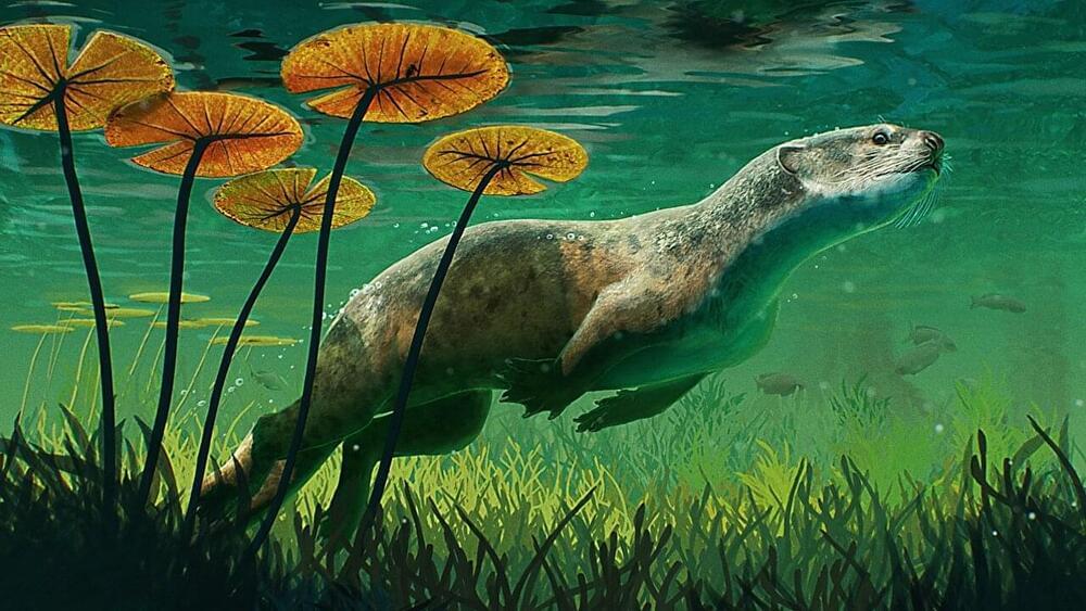
Toggle light / dark theme

Two Halves of the Hippocampus Have Different Gene Activity

Summary: Researchers have identified significant differences in gene activity between the anterior and posterior areas of the hippocampus. Genes associated with depression and other mood disorders are more active in the anterior hippocampus, while genes linked to cognitive disorders, such as ASD, are more active in the posterior hippocampus.

Source: UT Southwestern Medical Center.

A study of gene activity in the brain’s hippocampus, led by UT Southwestern researchers, has identified marked differences between the region’s anterior and posterior portions.

Dr. Matt Kaeberlein — Founder / Co-Director, Dog Aging Project — Professor, University of Washington

Enrolling Tens Of Thousands Of Dogs, In 10-Year Study, To Unlock Healthy Aging Secrets — Dr Matt Kaeberlein, Founder / Co-Director, The Dog Aging Project, Professor, University of Washington, joins me on Progress, Potential, And Possibilities Nathan Shock Centers #Rapamycin #Dogs #Aging #Longevity #Healthspan #Geroscience.

#MitochondrialDisease


Dr. Matt Kaeberlein is Professor of Pathology, Adjunct Professor of Genome Sciences, and Adjunct Professor of Oral Health Sciences, at the University of Washington.

Dr. Kaeberlein received his PhD from MIT in Biology, did his post-doc in the Department of Genome Sciences, University of Washington, and his research interests are focused on basic mechanisms of aging in order to facilitate translational interventions that promote healthspan and improve quality of life.

Dr. Kaeberlein has published nearly 200 papers in top scientific journals and has been recognized by several prestigious awards, including a Breakthroughs in Gerontology Award, an Alzheimer’s Association Young Investigator Award, an Ellison Medical Foundation New Scholar in Aging Award, a Murdock Trust Award, a Pioneer in Aging Award, and the Vincent Cristofalo Rising Star in Aging Research.

‘Amazing Natural Experiment’: In This Amazonian Tribe, Brains Don’t Age Like Ours

The Tsimane, an indigenous people who live in the Bolivian peripheries of the Amazon rainforest, lead lives that are very different to ours. They seem to be much healthier for it.

This tribal and largely isolated population of forager-horticulturalists still lives today by traditional ways of farming, hunting, gathering, and fishing – continuing the practices of their ancestors, established in a time long before industrialization and urbanization transformed most of the world.

For the Tsimane, the advantages are considerable. A study published in 2017 found that they effectively have the healthiest hearts in the world, with the lowest reported levels of coronary artery disease of any population ever recorded.

Computer simulations of the brain can predict language recovery in stroke survivors

At Boston University, a team of researchers is working to better understand how language and speech is processed in the brain, and how to best rehabilitate people who have lost their ability to communicate due to brain damage caused by a stroke, trauma, or another type of brain injury. This type of language loss is called aphasia, a long-term neurological disorder caused by damage to the part of the brain responsible for language production and processing that impacts over a million people in the US.

“It’s a huge problem,” says Swathi Kiran, director of BU’s Aphasia Research Lab, and College of Health & Rehabilitation Sciences: Sargent College associate dean for research and James and Cecilia Tse Ying Professor in Neurorehabilitation. “It’s something our lab is working to tackle at multiple levels.”

For the last decade, Kiran and her team have studied the brain to see how it changes as people’s improve with speech . More recently, they’ve developed new methods to predict a person’s ability to improve even before they start therapy. In a new paper published in Scientific Reports, Kiran and collaborators at BU and the University of Texas at Austin report they can predict recovery in Hispanic patients who speak both English and Spanish fluently—a group of aphasia patients particularly at risk of long-term language loss—using sophisticated computer models of the brain. They say the breakthrough could be a game changer for the field of speech therapy and for stroke survivors impacted by aphasia.

Expression of human‐specific ARHGAP11B in mice leads to neocortex expansion and increased memory flexibility

Generation of an ARHGAP11B-transgenic mouse line.

To generate a stable transgenic mouse line that expresses an ARHGAP11B protein, we first determined the temporal and spatial expression patterns of human ARHGAP11A and human ARHGAP11B mRNAs by qPCR of foetal human neocortical tissue at various developmental stages (gestational weeks 12–21; Fig EV1A and B) and by analysing previously published RNA-seq data sets of defined isolated NPC and neuron populations (Florio et al, 2015) (Fig EV1C and D), respectively. As the expression patterns of ARHGAP11A and ARHGAP11B mRNAs were found to be similar, we decided to generate the transgenic mouse line by converting one allele of the mouse Arhgap11a gene into a mutant mouse ARHGAP11B gene (mARHGAP11B), using the CRISPR/Cas9 genome editing technology (for details, see Materials and Methods). In m ARHGAP11B, the 55 nucleotides of Arhgap11a that in humans would be deleted from the ARHGAP11B mRNA by splicing using the new splice-donor site are replaced by the 141 nucleotides encoding the human-specific 47-amino acid sequence plus three nucleotides to generate a translational stop codon (Fig EV1E). Unless indicated otherwise, the ARHGAP11B-transgenic mice obtained (referred to as 11B mice hereafter) were used as heterozygous animals, that is, with one mouse Arhgap11a allele being replaced by m ARHGAP11B. The resulting ARHGAP11B protein will be expressed in developing mouse neocortex under the control of the native mouse Arhgap11a promotor.

Sonothermogenetics for noninvasive and cell-type specific deep brain neuromodulation

Critical advances in the investigation of brain functions and treatment of brain disorders are hindered by our inability to selectively target neurons in a noninvasive manner in the deep brain.

This study aimed to develop sonothermogenetics for noninvasive, deep-penetrating, and cell-type-specific neuromodulation by combining a thermosensitive ion channel TRPV1 with focused ultrasound (FUS)-induced brief, non-noxious thermal effect.

The sensitivity of TRPV1 to FUS sonication was evaluated in vitro. It was followed by in vivo assessment of sonothermogenetics in the activation of genetically defined neurons in the mouse brain by two-photon calcium imaging. Behavioral response evoked by sonothermogenetic stimulation at a deep brain target was recorded in freely moving mice. Immunohistochemistry staining of ex vivo brain slices was performed to evaluate the safety of FUS sonication.

A Massive New Gene Editing Project Is Out to Crush Alzheimer’s

The idea is simple: decades of research have found certain genes that seem to increase the chance of Alzheimer’s and other dementias. The numbers range over hundreds. Figuring out how each connects or influences another—if at all—takes years of research in individual labs. What if scientists unite, tap into a shared resource, and collectively solve the case of why Alzheimer’s occurs in the first place?

The initiative’s secret weapon is induced pluripotent stem cells, or iPSCs. Similar to most stem cells, they have the ability to transform into anything—a cellular genie, if you will. iPSCs are reborn from regular adult cells, such as skin cells. When transformed into a brain cell, however, they carry the original genes of their donor, meaning that they harbor the original person’s genetic legacy—for example, his or her chance of developing Alzheimer’s in the first place. What if we introduce Alzheimer’s-related genes into these reborn stem cells, and watch how they behave?

By studying these iPSCs, we might be able to follow clues that lead to the genetic causes of Alzheimer’s and other dementias—paving the road for gene therapies to nip them in the bud.

Neuralink Brain Chip Will End Language in Five to 10 Years, Elon Musk Says

In a recent interview, Elon Musk stated that the human language could possibly end within five to ten years. The CEO of Neuralink went to talk with Joe Rogan, implying that with the innovation of the brain chip the company is currently developing, humans won’t have to speak anymore using traditional languages.


Neuralink develops a chip that will soon be able to attach to the human brain. The chip’s invention aimed to communicate faster and conveniently. Through a single universal language, Elon Musk believes that the way we talk today will soon improve. The brain chip is expected to be completed to be developed within a few years, and by then, our communication could possibly evolve.

Elon Musk stated that the Neuralink chip’s success may take a while, but it should take five to ten years if the development will accelerate. He also added that the progress of the brain chip is on track, but with only to focus on their current objective, which is to help people minimize and prevent brain injuries, Express reported.

Elon Musk’s Brain Chip Completion on Progress, Initial Batch Will Solve Brain Injuries

The brain chip’s power was already displayed in a 2019 long-stream video by Neuralink. In the video published on the YouTube channel Monkey MindPong, a monkey with an active chip on its brain was shown playing a video game that is similar to a table tennis match. The astounding display of the monkey’s brain reaction toward the game left experts in awe.