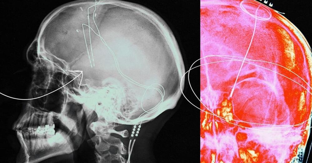
A team of researchers at the University of Massachusetts Amherst has developed the first dual-color optoelectronic neural probe.
Unlike previous, single probes, which often control brain activity in only one direction—excitation or inhibition, but not both—this new design can enhance and silence the electrical activities of the same neurons within specific cortical layers of the brain. It promises aid the investigation of tightly packed neural microcircuits within the cortex and deep brain regions and, in the longer term, add to the functional mapping of the brain.
Guangyu Xu, assistant associate professor of electrical and computer engineering, an appointee of the Dev and Linda Gupta Professorship at UMass Amherst, and principal investigator of the study hopes the device can ultimately help researchers identify the origin of brain diseases.
For the first time ever, deep brain stimulation has been shown to help people regain cognitive abilities they’d lost following a traumatic brain injury (TBI):
The lasting symptoms of a traumatic brain injury (TBI) can be treated with deep brain stimulation, according to a first-of-its kind trial.
For many people struggling with obesity, the drug is a potential lifesaver. Excess weight is associated with higher incidences of stroke, heart and liver disease, sleep apnea, joint problems, and some cancers. A major clinical trial this year in tens of thousands of overweight people without diabetes found the main ingredient in Ozempic, semaglutide, reduced the risk of stroke and heart attack, while lowering the chances of death due to cardiovascular problems.
Perhaps even more importantly, the drug is gradually changing societal views on obesity—it’s not due to lack of will power, but a chronic medical condition that can be treated.
But Ozempic and similar drugs—like Wegovy, another semaglutide-based medication that has been FDA-approved for weight loss—are already set for the next chapter: tackling a wide range of brain disorders, including Alzheimer’s and Parkinson’s. Clinical trials are underway for addiction, and the drugs are showing early promise battling bipolar disorder and depression.
But what would it actually mean to transfer your mind from “meat space” to cyberspace, and how could it be done? The basic idea rests on several assumptions, says Angela Thornton, a researcher at the Horizon Centre for Doctoral Training at University of Nottingham, who is also partnered with the Carboncopies Foundation, a non-profit that focuses on “whole brain emulation” and the creation of substrate-independent minds. “It assumes that we could replicate our brain [with] a certain level of understanding of how it works,” she says. “Not necessarily knowing all the detail, but enough to be able to emulate it.” Then, she adds, we have to make the assumption that the “mind” (i.e. the abstract part of us that thinks, remembers, imagines and senses) naturally emerges from the structures of the physical brain.
This is a lot to take on, which is partly why current brain emulation research is still stuck at the level of worms and, in more advanced studies, mice. Whether you agree with them or not, though, the arguments to take experiments further – toward larger mammals and, finally, humans – are quite obvious. For one, we could theoretically ‘live’ forever as a disembodied consciousness (or at least until the machines that hosted our virtual minds were destroyed), and continue interacting with our loved ones after they’ve passed as well. It’s possible that this could also go some way to solving the alleged population crisis, while limiting the impact of our physical bodies on the planet’s finite resources.
Of course, there are plenty of important questions that need answers before any of this can actually happen. Below, Thornton helps us unpick some of the main constraints and controversies.
Optogenetics has revolutionized neuroscience understanding by allowing spatiotemporal control over cell-type specific neurons in neural circuits. However, the sluggish development of noninvasive photon delivery in the brain has limited the clinical application of optogenetics. Focused ultrasound (FUS)-derived mechanoluminescence has emerged as a promising tool for in situ photon emission, but there is not yet a biocompatible liquid-phase mechanoluminescence system for spatiotemporal optogenetics. To achieve noninvasive optogenetics with a high temporal resolution and desirable biocompatibility, we have developed liposome (Lipo@IR780/L012) nanoparticles for FUS-triggered mechanoluminescence in brain photon delivery. Synchronized and stable blue light emission was generated in solution under FUS irradiation due to the cascade reactions in liposomes.
Humans and other animals with brains perhaps aren’t the only beings on the planet to experience consciousness, says a study in the journal EMBO Reports.
Consciousness instead underpins all life forms, from the smallest cells to the most complex organisms. Far from being limited to creatures like ourselves, the cell-based theory of consciousness frames the phenomenon a fundamental part of life itself.
Conventional thinking about consciousness—called the standard model of consciousness—focuses on the brain, supposing only complex organisms like humans and animals have it. But the new cell-based theory argues that consciousness started with the very first cells that emerged about 3.8 billion years ago and plants, bacteria and even amoebas have it.
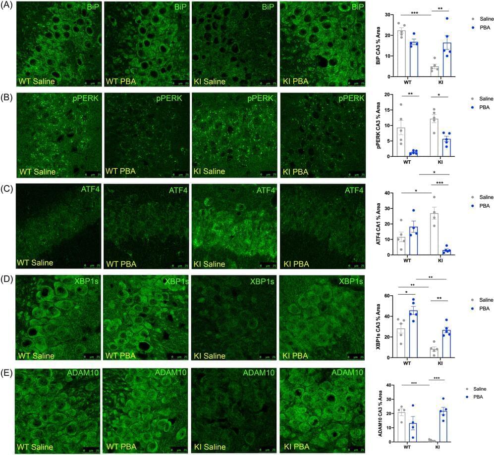
A “chaperone” molecule that slows the formation of certain proteins reversed disease signs, including memory impairment, in a mouse model of Alzheimer’s disease, according to a study from researchers at the Perelman School of Medicine at the University of Pennsylvania.
In the study, published in Aging Biology, researchers examined the effects of a compound called 4-phenylbutyrate (PBA), a fatty-acid molecule known to work as a “chemical chaperone” that inhibits protein accumulation. In mice that model Alzheimer’s disease, injections of PBA helped to restore signs of normal proteostasis (the protein regulation process) in the animals’ brains while also dramatically improving their performance on a standard memory test, even when administered late in the disease course.
“By generally improving neuronal and cellular health, we can mitigate or delay disease progression,” said study senior author Nirinjini Naidoo, Ph.D., a research associate professor of Sleep Medicine. “In addition, reducing proteotoxicity— irreparable damage to the cell that is caused by an accumulation of impaired and misfolded proteins—can help improve some previously lost brain functions.”
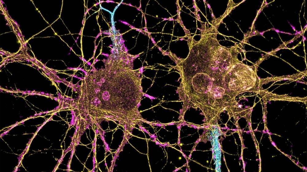
Neurons communicate through chemical signals known as neurotransmitters. Researchers at St. Jude Children’s Research Hospital, leveraging their expertise in structural biology, have successfully elucidated the structures of the vesicular monoamine transporter 2 (VMAT2), a key component of neuronal communication.
By visualizing VMAT2 in different states, scientists now better understand how it functions and how the different shapes the protein takes influence drug binding — critical information for drug development to treat hyperkinetic (excess movement) disorders such as Tourette syndrome. The work was recently published in the journal Nature.

