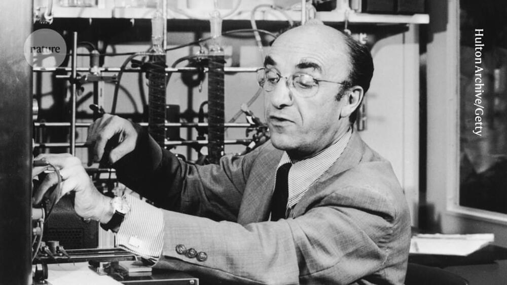
Toggle light / dark theme

Researchers decode patterns that make our brains human

The human brain may be the most complex piece of organized matter in the known universe, but Allen Institute researchers have begun to unravel the genetic code underlying its function. Research published this month in Nature Neuroscience identified a surprisingly small set of molecular patterns that dominate gene expression in the human brain and appear to be common to all individuals, providing key insights into the core of the genetic code that makes our brains distinctly human.

“So much research focuses on the variations between individuals, but we turned that question on its head to ask, what makes us similar?” says Ed Lein, Ph.D., Investigator at the Allen Institute for Brain Science. “What is the conserved element among all of us that must give rise to our unique cognitive abilities and human traits?”

Researchers used data from the publicly available Allen Human Brain Atlas to investigate how gene expression varies across hundreds of functionally distinct brain regions in six human brains. They began by ranking genes by the consistency of their expression patterns across individuals, and then analyzed the relationship of these genes to one another and to brain function and association with disease.

Two heads are better than one: Gene expression reveals molecular mechanisms underlying evolution of cerebral cortex

Dramatic expansion of the human cerebral cortex, over the course of evolution, accommodated new areas for specialized cognitive function, including language. Understanding the genetic mechanisms underlying these changes, however, remains a challenge to neuroscientists.

A team of researchers in Japan has now elucidated the mechanisms of cortical evolution. They used molecular techniques to compare the in mouse and monkey brains.

Using the technique called in situ hybridization to visualize the distribution of mRNA transcripts, Okano, Shimogori and their colleagues examined the expression patterns of genes that are known to regulate development of the . They compared these patterns to those of the same genes in the brain of the common marmoset. They found that most of the genes had similar expression patterns in mice and marmosets, but that some had strikingly different patterns between the two species. Notably, some areas of the visual and prefrontal showed expression patterns that were unique to marmosets.

The beginnings of the thinking brain

Oxford researchers have identified the very first neurons in the human cerebral cortex, the part of the brain that sets us apart from all other animals.

Dr Irina Bystron and colleagues from the Department of Physiology, Anatomy and Genetics at the University of Oxford, together with Professor Pasko Rakic, a leading neuroscientist at Yale University, describe for the first time in Nature Neuroscience the very earliest nerve cells in the part of the developing human brain that becomes the cerebral cortex.

The cerebral cortex is largely responsible for human cognition, playing an essential role in perception, memory, thought, language, mental ability, intellect and consciousness. It is also responsible for our voluntary actions. As adults our cerebral cortex accounts for 40 per cent of the brain’s weight and is composed of about 20 billion neurons. The new findings show that its first neurons are in place much earlier than previously thought – approximately 31 days after fertilization, when the entire embryo is only about 4 mm long and shaped a bit like a comma, before the development of arms, legs or eyes.

Biochemists use enzymes to change how brain cells communicate with each other

As you’re reading this sentence, the cells in your brain, called neurons, are sending rapid-fire electrical signals between each other, transmitting information. They’re doing so via tiny, specialized junctions between them called synapses.

There are many different types of that form between neurons, including “excitatory” or “inhibitory,” and the exact mechanisms by which these structures are generated remain unclear to scientists. A Colorado State University biochemistry lab has uncovered a major insight into this question by showing that the types of chemicals released from synapses ultimately guide which kinds of synapses form between neurons.

Soham Chanda, assistant professor in the Department of Biochemistry and Molecular Biology, led the study published in Nature Communications that demonstrates the possibility of changing the identity of synapses between neurons, both in vitro and in vivo, through enzymatic means. The other senior scientists who contributed to the project were Thomas Südhof of Stanford University and Matthew Xu-Friedman of the University at Buffalo.

How Dopamine Plays a Key Role in Consciousness

Summary: Study reveals how dopamine may have a central role in maintaining our consciousness.

Source: The Conversation.

Consciousness is arguably the most important scientific topic there is. Without consciousness, there would after all be no science. But while we all know what it is like to be conscious – meaning that we have personal awareness and respond to the world around us – it has turned out to be near impossible to explain exactly how it arises from the hardware of the brain. This is dubbed the “hard” problem of consciousness.

Awareness Without a Sense of Self

Summary: People who practice meditation often report feeling “pure awareness” in which they say they experience consciousness itself. The state encompasses specific sensations and non-specific feelings, thoughts, and perceptions. Researchers say their findings will help explain “pure consciousness,” and work to generate a prototypical minimal model for human conscious perception.

Source: Johannes Gutenberg University Mainz.

In the context of meditation practice, meditators can experience a state of “pure awareness” or “pure consciousness”, in which they perceive consciousness itself. This state can be experienced in various ways, but evidently incorporates specific sensations as well as non-specific accompanying perceptions, feelings, and thoughts.

Antibiotic Treatments May Make Us More Susceptible to Negative Emotions

Summary: Recent antibiotic use impacts the way in which people pay attention to negative facial expressions. Findings shed light on how antibiotic use can increase the risks of depression.

Source: Lieden University.

People who have taken antibiotics in the past three months pay more attention to negative facial expressions, according to research by postdoc Katerina Johnson and assistant professor Laura Steenbergen. This may explain how antibiotics increase the risk of developing depression.

Neural prosthetics: Krishna Shenoy at TEDxStanford

Krishna Shenoy helps to restore lost function for disabled patients by designing prosthetic devices that can translate neural brain activity.

Krishna Shenoy directs the Neural Prosthetic Systems Lab, where his group conducts neuroscience and neuro-engineering research to better understand how the brain controls movement and to design medical systems to assist those with movement disabilities. Shenoy also co-directs the Neural Prosthetics Translational Lab, which uses these advances to help people with severe motor disabilities. Shenoy received his bachelor’s degree in electrical engineering from UC-Irvine and his master’s and doctoral degrees in the same field from MIT. He was a neurobiology postdoctoral fellow at Caltech in Pasadena and then joined Stanford University, where he is a professor of electrical engineering, bioengineering and neurobiology.

What Does GABA Do in the Brain?

Despite the fact that sex is a basic instinct and a near-universal experience, we know remarkably little about it. And so, this week, we’re teaming up with our friends at Futurism, oracles of all things science, technology and medicine, to look at the past, present and future of pleasure from a completely scientific perspective.

For a while now, the neurotransmitter dopamine has been seen as the conductor of good feelings. It’s the subject of love songs, the seductress of biohackers and the ostensible “pleasure chemical.” But as research continues to uncover more about our brain’s reward system, dopamine is beginning to look less like the maestro and more like a member of the band.

Say Goodbye to Binge Eating: Signal Pathway in Brain That Controls Food Intake Discovered

A group of scientists has developed an entirely new approach to treating eating disorders.

They showed that a group of nerve cells (so-called AgRP, agouti-related peptide neurons) in the hypothalamus control the release of endogenous lysophospholipids, which in turn control the excitability of nerve cells in the cerebral cortex, which stimulates food intake.

In this process, the crucial step of the signaling pathway is controlled by autotaxin, an enzyme that is responsible for the production of lysophosphatidic acid.