Year 2013 face_with_colon_three
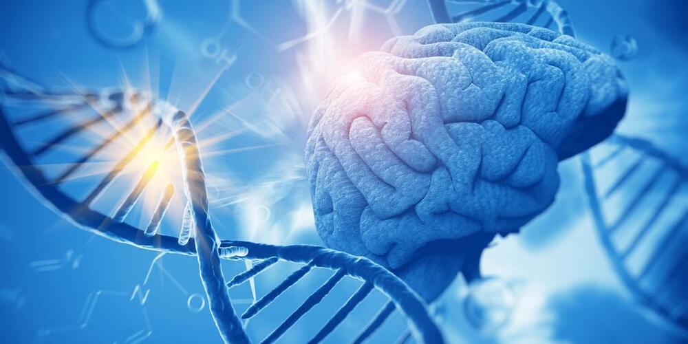
Scientists at Oregon Health & Science University and the Oregon National Primate Research Center (ONPRC) have successfully reprogrammed human skin cells to become embryonic stem cells capable of transforming into any other cell type in the body. It is believed that stem cell therapies hold the promise of replacing cells damaged through injury or illness. Diseases or conditions that might be treated through stem cell therapy include Parkinson’s disease, multiple sclerosis, cardiac disease and spinal cord injuries.
The research breakthrough, led by Shoukhrat Mitalipov, Ph.D., a senior scientist at ONPRC, follows previous success in transforming monkey skin cells into embryonic stem cells in 2007. This latest research will be published in the journal Cell online May 15 and in print June 6.
The technique used by Drs. Mitalipov, Paula Amato, M.D., and their colleagues in OHSU’s Division of Reproductive Endocrinology and Infertility, Department of Obstetrics & Gynecology, is a variation of a commonly used method called somatic cell nuclear transfer, or SCNT. It involves transplanting the nucleus of one cell, containing an individual’s DNA, into an egg cell that has had its genetic material removed. The unfertilized egg cell then develops and eventually produces stem cells.