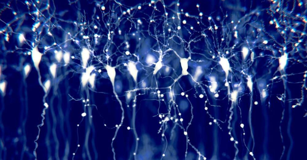
The Allen Institute’s release includes recordings from a whopping 300,000 mouse neurons. Now the challenge is figuring out what to do with all that data.

Neuroscience is contributing to an understanding of the biological bases of human intelligence differences. This work is principally being conducted along two empirical fronts: genetics—quantitative and molecular—and brain imaging. Quantitative genetic studies have established that there are additive genetic contributions to different aspects of cognitive ability—especially general intelligence—and how they change through the lifespan. Molecular genetic studies have yet to identify reliably reproducible contributions from individual genes. Structural and functional brain-imaging studies have identified differences in brain pathways, especially parieto-frontal pathways, that contribute to intelligence differences. There is also evidence that brain efficiency correlates positively with intelligence.

In 1906, Alois Alzheimer, a psychiatrist and neuroanatomist, reported “a peculiar severe disease process of the cerebral cortex” to a gathering of psychiatrists in Tübingen, Germany.
The case was a 50-year-old woman who suffered from memory loss, delusions, hallucinations, aggression, and confusion – all of which worsened until her untimely death five years later.
In the autopsy, Alzheimer noticed distinctive plaques on her brain. These plaques – clumps of amyloid-beta protein – are still considered to be the cause of Alzheimer’s disease.

In 1906, Alois Alzheimer, a psychiatrist and neuroanatomist, reported “a peculiar severe disease process of the cerebral cortex” to a gathering of psychiatrists in Tübingen, Germany. The case was a 50-year-old woman who suffered from memory loss, delusions, hallucinations, aggression and confusion – all of which worsened until her untimely death five years later.
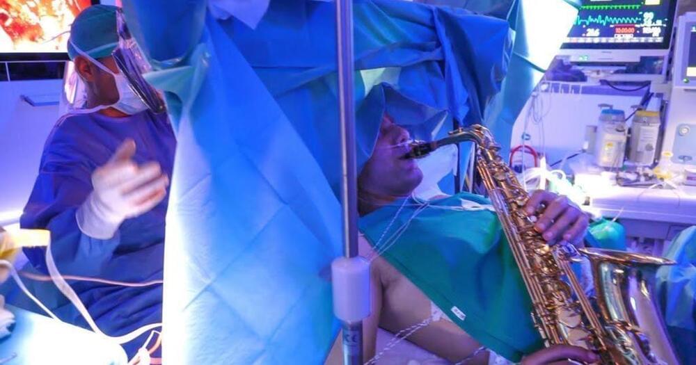
A dish of living brain cells has learned to play the 1970s arcade game Pong.
About 800,000 cells linked to a computer gradually learned to sense the position of the game’s electronic ball and control a virtual paddle, a team reports in the journal Neuron.
The novel achievement is part of an effort to understand how the brain learns, and how to make computers more intelligent.
O.o!!!.
Johns Hopkins researchers say that an experimental anticancer compound appears to have reversed behaviors associated with schizophrenia and restored some lost brain cell function in adolescent mice with a rodent version of the devastating mental illness.
The drug is one of a class of compounds known as PAK inhibitors, which have been shown in animal experiments to confer some protection from brain damage due to Fragile X syndrome, an inherited disease in humans marked by mental retardation. There also is some evidence, experts say, suggesting PAK inhibitors could be used to treat Alzheimer’s disease. And because the PAK protein itself can initiate cancer and cell growth, PAK inhibitors have also been tested for cancer.
In the new Johns Hopkins-led study, reported online March 31 in the Proceedings of the National Academy of Sciences, the researchers found that the compound, called FRAX486, appears to halt an out-of-control biological “pruning” process in the schizophrenic brain during which important neural connections are unnecessarily destroyed.

Circa 2018 face_with_colon_three
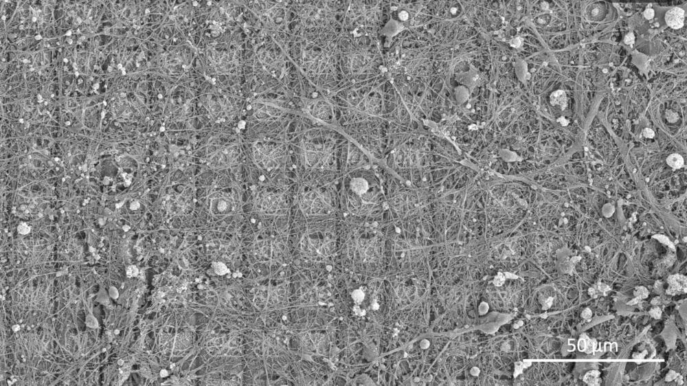
Since the time of Hippocrates and Herophilus, scientists have placed the location of the mind, emotions and intelligence in the brain. For centuries, this theory was explored through anatomical dissection, as the early neuroscientists named and proposed functions for the various sections of this unusual organ. It wasn’t until the late 19th century that Camillo Golgi and Santiago Ramón y Cajal developed the methods to look deeper into the brain, using a silver stain to detect the long, stringy cells now known as neurons and their connections, called synapses.
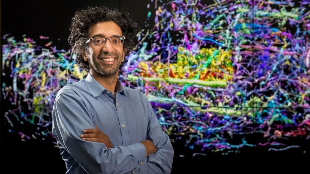
Today, neuroanatomy involves the most powerful microscopes and computers on the planet. Viewing synapses, which are only nanometers in length, requires an electron microscope imaging a slice of brain thousands of times thinner than a sheet of paper. To map an entire human brain would require 300,000 of these images, and even reconstructing a small three-dimensional brain region from these snapshots requires roughly the same supercomputing power it takes to run an astronomy simulation of the universe.
Fortunately, both of these resources exist at Argonne, where, in 2015, Kasthuri was the first neuroscientist ever hired by the U.S. Department of Energy laboratory. Peter Littlewood, the former director of Argonne who brought him in, recognized that connectome research was going to be one of the great big data challenges of the coming decades, one that UChicago and Argonne were perfectly poised to tackle.
Circa 2017 face_with_colon_three
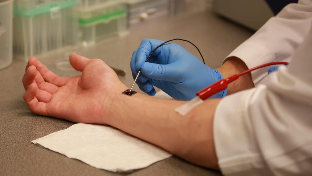
A new device developed at The Ohio State University can start healing organs in a “fraction of a second,” researchers say.
The technology, known as Tissue Nanotransfection (TNT), has the potential to save the lives of car crash victims and even deployed soldiers injured on site. It’s a dime-sized silicone chip that “injects genetic code into skin cells, turning those skin cells into other types of cells required for treating diseased conditions,” according to a release.
In lab tests, one touch of TNT completely repaired injured legs of mice over three weeks by turning skin cells into vascular cells.