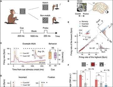
View all available purchase options and get full access to this article.
Citation information is sourced from Crossref Cited-by service.
A neurosurgeon in Australia pulled a wriggling 3-inch roundworm from the brain of a 64-year-old woman last year—which was quite the surprise to the woman’s team of doctors and infectious disease experts, who had spent over a year trying to identify the cause of her recurring and varied symptoms.
A close study of the extracted worm made clear why the diagnosis was so hard to pin down: the roundworm was one known to infect snakes—specifically carpet pythons endemic to the area where the woman lived—as well as the pythons’ mammalian prey. The woman is thought to be the first reported human to ever have an infection with this snake-adapted worm, and it is the first time the worm has been found burrowing through a mammalian brain.
When the woman’s illness began, “trying to identify the microscopic larvae, which had never previously been identified as causing human infection, was a bit like trying to find a needle in a haystack,” Karina Kennedy, a professor at the Australian National University (ANU) Medical School and Director of Clinical Microbiology at Canberra Hospital, said in a press release.
Sepideh Sadaghiani, Associate Professor of Psychology, Neuroscience, & Bioengineering at Illinois, lectured on “The functional connectome across temporal scales” at 4:00 pm in 2,269 Beckman Institute and on Zoom. Introduction by Ryan Miller, MBM trainee and PhD candidate in Chemical & Biomolecular Engineering.
For more information on the lecture and Dr. Sadaghiani: https://publish.illinois.edu/minibrain/2022/07/26/sepideh-sa…s-lecture/
For a list of our upcoming events: https://minibrain.beckman.illinois.edu/events/
This video was supported by the National Science Foundation under grant 1735252. https://minibrain.beckman.illinois.edu/
Plenary Talk by Michael Levin on “Non-neural, developmental bioelectricity as a precursor for cognition: Evolution, synthetic organisms, and biomedicine” at the Virtual Miniature Brain Machinery Retreat, September 16, 2021. Introduction by William Baker.
Michael Levin.
Director of the Allen Discovery Center.
Tufts University.
Sponsored by the National Science Foundation, the University of Illinois at Urbana-Champaign, and the Beckman Institute for Advanced Science & Technology. This video was supported by the National Science Foundation under grant 1735252.
Being able to vocalize is one of the most essential elements of the human experience, with infants expected to start babbling their first words before they’re one year old, and much of their further life revolving around interacting with others using vocalizations involving varying degrees of vocabulary and fluency. This makes the impairment or loss of this ability difficult to devastating, as is the case with locked-in syndrome (LIS), amyotrophic lateral sclerosis (ALS) and similar conditions, where talking and vocalizing has or will become impossible.
In a number of concurrent studies, the use of a brain-computer interface (BCI) is investigated to help patients suffering from LIS (Sean L. Metzger et al., 2023) and ALS (Francis R. Willett et al., 2023) to regain their speaking voice. Using the surgically implanted microelectrode arrays (Utah arrays) electrical impulses pertaining to the patient’s muscles involved in speaking are recorded and mapped to phonemes, which are the elements that make up speech. Each of these phonemes requires a specific configuration of the muscles of the vocal tract (e.g. lips, tongue, jaw and larynx), which can be measured with a fair degree of accuracy.
In the case of the study by Sean L. Metzger et al. as recently published in Nature, the accompanying research article on the University of California San Francisco website details the story of their patient: Ann. At the age of 30, Ann suffered a brainstem stroke which rendered her essentially fully paralyzed. As an LIS patient she lacked for a long time even the ability to move her facial muscles.
A massive study of medical and genetic data shows that people with a particular version of a gene involved in immune response had a lower risk of Alzheimer’s and Parkinson’s disease.
Specific changes in our DNA that increase the risk of developing epilepsy have been discovered, in the largest genetic study of its kind for epilepsy coordinated by the International League Against Epilepsy, which includes scientists from the University of Melbourne and WEHI (Walter and Eliza Hall Institute of Medical Research).
Published today in Nature Genetics, this research advances our understanding of why epilepsy develops and could inform the development of new epilepsy treatments. The research was produced by the International League Against Epilepsy (ILAE) Consortium on Complex Epilepsies.
Epilepsy is a common brain disorder estimated to effect more than 50 million people worldwide, where nerve cell activity in the brain is disturbed, causing seizures. It has a genetic component that sometimes runs in families. In this study, researchers compared the DNA from almost 30,000 people with epilepsy to the DNA of 52,500 people without epilepsy from around the world. The differences between the two groups highlighted areas of DNA that may be involved in the development of epilepsy.
A neonatal hypoxic-injury animal model revealed that CK2α mediated Daam2 phosphorylation, which plays a protective role in developmental and behavioral recovery after neonatal hypoxia, a form of brain injury seen in cerebral palsy and other conditions. Additionally, it facilitates remyelination after white matter injury in adult animals.
Together, these findings have identified a novel regulatory node connecting CK2α and Daam2 in the Wnt pathway that regulates stage-specific oligodendrocyte development and offers insights into a new biological mechanism to regenerate myelin.
“This study opens exciting therapeutic avenues we could develop in the future to repair and restore myelin, which has the potential to alleviate and treat several neurological issues that are currently untreatable,” Lee said.
We live in an analog world of continuous information flow that is both processed and stored by our brains at the same time, but our devices process information digitally in the form of discrete binary code, breaking the information into little bits (or bites).
Researchers at EPFL have revealed a pioneering technology that combines the potential of continuous analog processing with the precision of digital devices. By seamlessly integrating ultra-thin, two-dimensional semiconductors with ferroelectric materials, the research, published in Nature Electronics, unveils a novel way to improve energy efficiency and add new functionalities in computing. The new configuration merges traditional digital logic with brain-like analog operations.
The innovation from the Nanoelectronics Device Laboratory (Nanolab), in collaboration with Microsystems Laboratory, revolves around a unique combination of materials leading to brain-inspired functions and advanced electronic switches, including the standout negative capacitance Tunnel Field-Effect Transistor (TFET).