Researchers created the most detailed map of the brain’s functional networks using data from people watching movies, including “Inception,” “Home Alone” and “Erin Brokovich.”


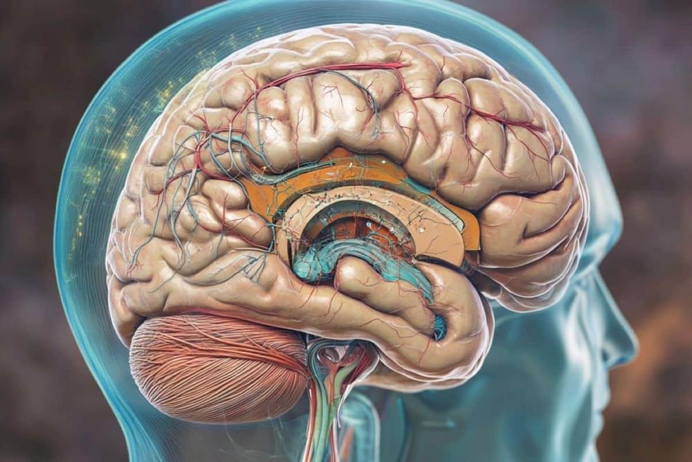
Johns Hopkins University-led researchers, working with the Biomarkers for Older Controls at Risk for Dementia (BIOCARD) cohort, have found that certain factors are linked to faster brain shrinkage and quicker progression from normal thinking abilities to mild cognitive impairment (MCI). People with type 2 diabetes and low levels of specific proteins in their cerebrospinal fluid showed more rapid brain changes and developed MCI sooner than others.
Long-term studies tracking brain changes over many years are rare but valuable. Previous research mostly provided snapshots in time, which can’t show how individual brains change over the years. By following participants for up to 27 years (20-year median), this study offers new insights into how health conditions might speed up brain aging.
In a study, “Acceleration of Brain Atrophy and Progression From Normal Cognition to Mild Cognitive Impairment,” published in JAMA Network Open, researchers used the BIOCARD cohort to examine risk factors associated with the acceleration of brain atrophy and progression from normal cognition to MCI. An Invited Commentary is also available.
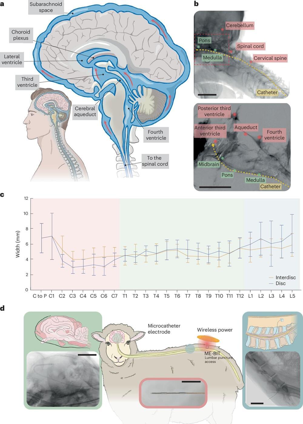
A team of researchers led by Rice University’s Jacob Robinson and the University of Texas Medical Branch’s Peter Kan has developed a technique for diagnosing, managing and treating neurological disorders with minimal surgical risks. The team’s findings were published in Nature Biomedical Engineering.
While traditional approaches for interfacing with the nervous system often require creating a hole in the skull to interface with the brain, the researchers have developed an innovative method known as endocisternal interfaces (ECI), allowing for electrical recording and stimulation of neural structures, including the brain and spinal cord, through cerebral spinal fluid (CSF).
“Using ECI, we can access multiple brain and spinal cord structures simultaneously without ever opening up the skull, reducing the risk of complications associated with traditional surgical techniques,” said Robinson, professor of electrical and computer engineering and bioengineering.
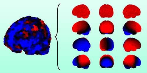
Summary: A recent study offers new insights into how brain regions coordinate during rest, using resting-state fMRI (rsfMRI) and neural recordings in mice. By comparing blood flow patterns with direct neural activity, researchers found that some brain activity remains “invisible” in traditional rsfMRI scans. This hidden activity suggests that current brain imaging techniques may miss key elements of neural behavior.
The findings, potentially applicable to human studies, may refine our understanding of brain networks. Further research could improve the accuracy of interpreting brain activity.
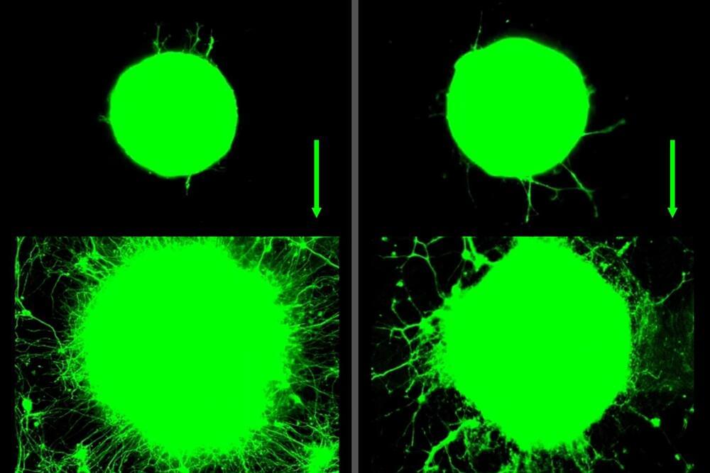
This study explores how muscle contractions, such as those that occur during exercise, influence motor neurons—the cells responsible for controlling muscle movement.
There’s no doubt that exercise does a body good. Regular activity not only strengthens muscles but can bolster our bones, blood vessels, and immune system.
Now, MIT engineers have found that exercise can also have benefits at the level of individual neurons. They observed that when muscles contract during exercise, they release a soup of biochemical signals called myokines.
In the presence of these muscle-generated signals, neurons grew four times further compared to neurons that were not exposed to myokines. These cellular-level experiments suggest that exercise can have a significant biochemical effect on nerve growth.


Satellite Data Reveals How Environment Shapes Kids’ Brain Health https://neurosciencenews.com/environment-brain-development-28026/
Adverse childhood experiences can lead to adult symptoms of anxiety and depression, mediated by life history strategies, according to a study published in Biodemography & Social Biology.
Existing research underscores the psychological impact of early-life adversity, with theories across cognitive, behavioral, and evolutionary psychology exploring these long-term effects. The life history theory, specifically, offers insight by framing adverse childhood experiences (ACEs) in terms of fast or slow life strategies, each with distinct reproductive and developmental adaptations suited to one’s environment.
Life history theory posits that individuals exposed to unstable or hostile environments in childhood often adopt a “fast” life strategy, focusing on early reproduction and risk-taking. In contrast, those in stable conditions tend to adopt “slow” strategies, emphasizing long-term planning and higher parental investment.