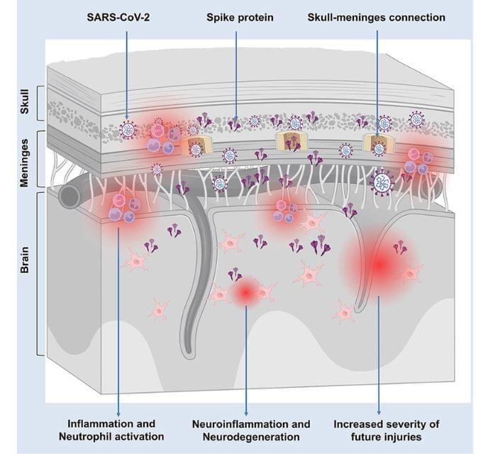
Toggle light / dark theme

Researchers at the University of Texas at Austin have developed a decoder that uses information from fMRI scans to reconstruct human thoughts. Jerry Tang, Amanda LeBel, Shailee Jain and Alexander Huth have published a paper describing their work on the preprint server bioRxiv.

Prior efforts to create technology that can monitor and decode them to reconstruct a person’s thoughts have all consisted of probes placed in the brains of willing patients. And while such technology has proven useful for research efforts, it is not practical for use in other applications such as helping people who have lost the ability to speak. In this new effort, the researchers have expanded on work from prior studies by applying findings about reading and interpreting brain waves to data obtained from fMRI scans.

Recognizing that attempting to reconstruct brainwaves into individual words using fMRI was impractical, the researchers designed a decoding device that sought to gain an overall understanding of what was going on in the mind rather than a word-for-word decoding. The decoder they built was a that accepted fMRI data and returned paragraphs describing general thoughts. To train their algorithm, the researchers asked two men and one woman to lie in an fMRI machine while they listened to podcasts and recordings of people telling stories.

This is the first study to record such electrical activity from inside the brain.

How do people remember the things they’ve learned? To get to the bottom of the mystery, scientists undertook a study that looked deep inside the brain.

Neuroscientists from Northwestern University and clinicians from the University of Chicago Epilepsy Center examined the electrical activity in the brains of five patients at the center in response to sounds administered by the research team as part of a learning exercise.


Department of neurological surgery, the university of chicago.

Recent evidence points out the role of the gut microbiota in the aging process. However, the specific changes and relevant interventions remain unclear. In this study, Senescence Accelerated Mouse-Prone 8 (SAMP8) mice were divided into four groups; young-FMT-group transplanted fecal microbiota from young donors (2–3°months old) and old-FMT-group transplanted from old donors (10–11°months old); additionally, other two groups either adult mice injected with saline solution or untreated mice served as the saline and blank control groups, respectively. All mice were intervened from their 7-months-old until 13-months-old. The open field test at 9 and 11°months of age showed that the mice transplanted with gut microbiota from young donors had significantly better locomotor and exploration ability than those of transplanted with old-donors gut microbiota and those of saline control while was comparable with the blank control. 16S rRNA gene sequencing showed that the gut microbiome of recipient mice of young donors was altered at 11°months of age, whereas the alternation of the gut microbiome of old-donor recipient mice was at 9°months. For comparison, the recipient mice in the blank and saline control groups exhibited changes in the gut microbiome at 10°months of age. The hallmark of aging-related gut microbiome change was an increase in the relative abundance of Akkermansia, which was significantly higher in the recipients transplanted with feces from older donors than younger donors at 9°months of age. This study shows that fecal microbiota transplantation from younger donors can delay aging-related declines in locomotor and exploration ability in mice by changing the gut microbiome.

Aging is inherently accompanied by the decline of physical and mental abilities, including locomotor, cognition, and bodily functions, to subsequently cause frailty syndrome, neurodegenerative diseases, and other age-related diseases, which reduce the quality of life of the aging population (Hou et al., 2019). Aging mechanisms and anti-aging interventions have long been a major focus of biomedical research, which is particularly relevant given the rapidly aging society.

The gut is a major organ for nutrients absorption, metabolism, and immunity, and contains hundreds of millions of microorganisms and their metabolites, which comprise the gut microbiota (Heintz and Mair, 2014) that interacts with host cells and tissues (Huang et al., 2021). Our previous study reported continuous changes in the gut microbiome of centenarians during their transition from a healthy status to death. The most significant changes of gut microbial communities in the period were found to occur at 7°months prior to death, suggesting that this may be a turning point of significant changes in the gut microbiome of centenarians (Luan et al., 2020). Recent studies have revealed an important relationship between the gut microbiome and aging-related diseases such as Alzheimer disease (Ticinesi et al., 2018; Haran and McCormick, 2021), suggesting that the gut microbiome plays an essential role in the aging process.

Fungi is getting stronger globally even alerting the WHO due to its damage.


The World Health Organization created a list of fungi that it said pose a growing risk to human health, including yeasts and molds found in abundance in nature and the body.

The WHO said Tuesday that the 19 species on the list merit urgent attention from public-health officials and drug developers. Four species were designated as threats of the highest priority: Aspergillus fumigatus, a mold found abundantly in nature; Candida albicans, which is commonly found in the human body; Candida auris, a highly deadly yeast; and Cryptococcus neoformans, a fungus that can cause deadly brain infections.

“Fungal infections are growing, and are ever more resistant to treatments, becoming a public-health concern worldwide” said Hanan Balkhy, the WHO’s assistant director-general.

Scientists can now “decode” people’s thoughts without even touching their heads, The Scientist reported.

Past mind-reading techniques relied on implanting electrodes deep in peoples’ brains. The new method, described in a report posted 29 Sept. to the preprint database bioRxiv, instead relies on a noninvasive brain scanning technique called functional magnetic resonance imaging (fMRI).

FMRI tracks the flow of oxygenated blood through the brain, and because active brain cells need more energy and oxygen, this information provides an indirect measure of brain activity.

Our immune system is the first line of defense against disease, but unfortunately it can go rogue and attack healthy tissues. Scientists at Johns Hopkins University have now engineered a protein that may help prevent these autoimmune diseases by boosting the number of regulatory T cells (Tregs).

The immune system keeps a vigilant watch over our bodies at all times, tagging and destroying foreign pathogens or problematic cells to prevent illness. However, sometimes it can get a little overzealous and start attacking the body’s own cells, which can trigger a range of autoimmune diseases like type 1 diabetes, lupus and rheumatoid arthritis.

To prevent these issues from arising, immune cells called Tregs play the vital role of keeping the immune system responses in check, but they can fail at this job. So for the new study, the researchers set out to boost their numbers, following previous studies that have shown promise in doing so to help treat autoimmune diseases like multiple sclerosis and Crohn’s disease.

Great ape animal studies have long been prohibited in Europe due to ethical concerns. An alternative to using animals in studies is the use of so-called organoids, which are three-dimensional cell structures that can be generated in the lab and are just a few millimeters in size.

These organoids can be created using pluripotent stem cells, which then subsequently develop into particular cell types like nerve cells. The study team was able to create both chimpanzee and human brain organoids by using this method.

“These brain organoids allowed us to investigate a central question concerning ARHGAP11B,” says Wieland Huttner of the Max Planck Institute of Molecular Cell Biology and Genetics, one of the three lead authors of the study.

Summary: A new 3D electrode array allows researchers to map the activity and location of up to 1 million synaptic links in a living brain.

Source: Rice University.

It’s a mystery how human thoughts and dreams emerge from electrical pulses in the brain’s estimated 100 trillion synapses, and Rice University neuroengineer Chong Xie dreams of changing that by creating a system that can record all the electrical activity in a living brain.