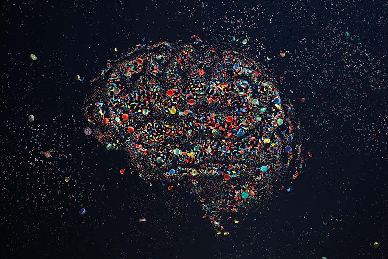
The average human brain contains around 7 grams of plastic, but it’s unclear how this affects us. Now animal studies are revealing links to poor cognition and weird behaviour
In this paper, I explore the limitations of various modern philosophical approaches to Chalmers’ Hard Problem of Consciousness, highlighting how the MVT/absent Primal Eye framework offers a compelling explanation for the qualitative nature of experience. We invite you to read these findings and share your thoughts on this intriguing intersection of philosophy and consciousness.
(https://www.academia.edu/144710257/Problems_with_modern_Philosophical_Solutions_to_Chalmer_s_Hard_Problem_of_Consciousness_and_how_the_loss_of_the_Primal_Eye_in_early_evolution_seemingly_gave_rise_to_waking_and_sleeping_Subjectivity)
Posthuman University Journal, October, 2025.
Materialist reductions fail to bridge the Explanatory Gap, functionalism fails to capture qualia, dissolutionism fails to account for phenomenal reality, and panpsychism collapses under the weight of the Combination Problem. The MVT/ absent Primal Eye philosophical framework seems to successfully explain the qualitative nature of experience without either denying its existence or creating an equally intractable set of metaphysical mysteries.
—
What they discovered.
Kampmann’s work, supported by the National Institutes of Science (NIH), maps cellular “decision points” that determine whether brain cells survive or die — laying the groundwork for treatments that intervene before irreversible brain damage occurs.
Using CRISPR-based gene targeting technology that his team helped develop and pioneer the use of in brain cells, Kampmann has identified genes and cellular processes that influence the buildup of amyloid plaque and tau in the brain, two primary contributors to dementia. Thanks to this technology, called CRISPR interference and CRISPR activation, select genes in the laboratory can be turned on or off to protect brain cells from decline.
“We can conduct large-scale experiments that target all the genes in the human genome — 20,000 of them,” said Kampmann, explaining his work after receiving the Byers Award earlier this year. “And that way, we can basically have a little knob on each gene to ask which of all of the genes play a role in a disease.”

People with autism spectrum disorders commonly have difficulty processing sensory information, which can make busy, bright or loud settings—such as schools, airports and restaurants—stressful or even painful. The neurological causes for altered sound processing are complex, and researchers are interested in better understanding them to make life better for people with autism.
In a study that combines behavioral tests, computer models and electrophysiological recordings of neuron activity, researchers have found that hyperactivity of neurons in the auditory cortex and the reaction of these neurons to an unusually broad range of frequencies contribute to this altered sound processing in rat models. The research is published in the journal PLOS Biology.
“One of the things we thought wasn’t being looked at enough was this idea of sensory discrimination: being able to distinguish between different features in our environment,” said Benjamin Auerbach, a professor of molecular and integrative physiology at the University of Illinois Urbana-Champaign.
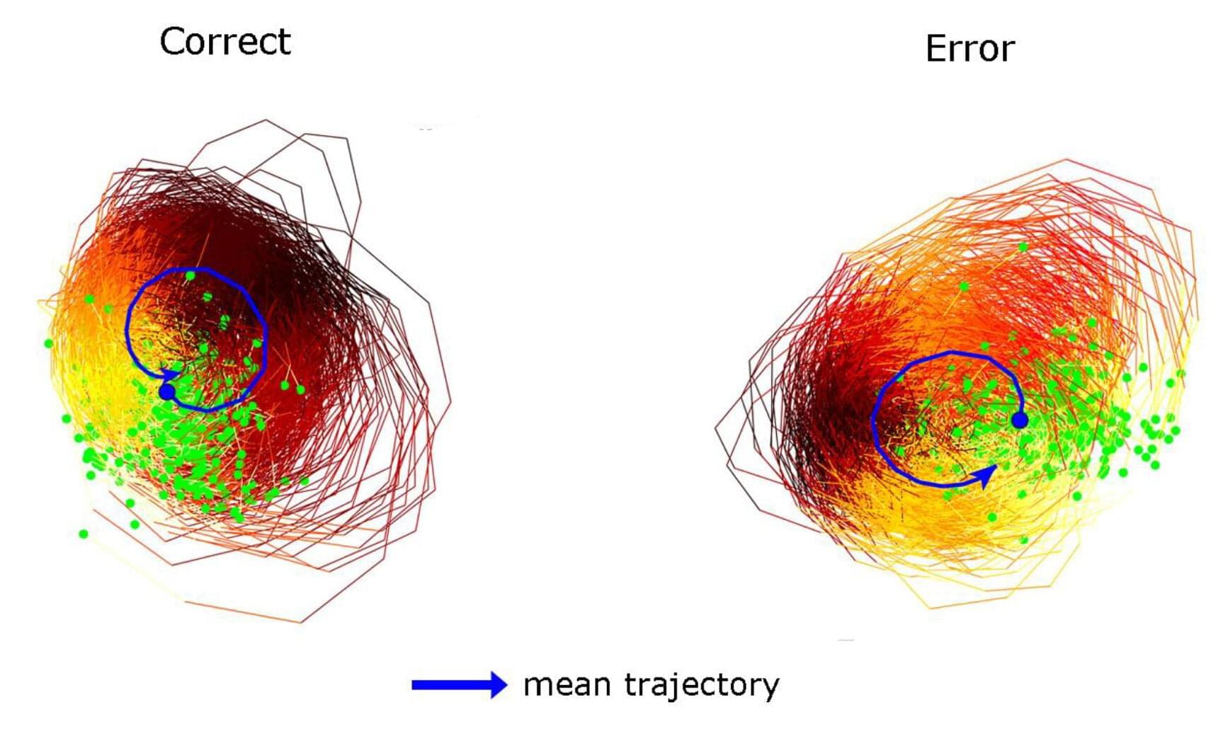
As sure as the brain is prone to distraction, it can also return its focus to the task at hand. A new study in animals by scientists at the Picower Institute for Learning and Memory of MIT shows how that seems to happen: Coordinated neural activity in the form of a rotating brain wave puts thought back on track.
“The rotating waves act like herders that steer the cortex back to the correct computational path,” said study senior author Earl K. Miller, Picower Professor in the Picower Institute and MIT’s Department of Brain and Cognitive Sciences.
Picower Institute postdoc Tamal Batabyal is the lead author of the study published in the Journal of Cognitive Neuroscience.
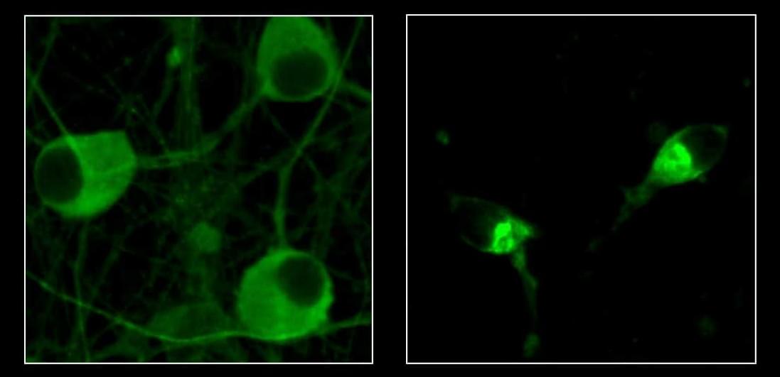
Researchers at the VIB-UAntwerp Center for Molecular Neurology have visualized how brain network development is altered in a model of KCNQ2-related developmental and epileptic encephalopathy, a rare childhood brain disorder. Using longitudinal imaging techniques, the team observed differences in how brain regions communicate and connect, long before behavioral symptoms appear.
KCNQ2-related developmental and epileptic encephalopathy (KCNQ2-DEE) is a rare but severe neurological disorder that affects newborns. Children with this condition typically develop seizures within days after birth and continue to face learning and movement difficulties. The disorder is caused by mutations in a potassium-channel gene that disrupts normal brain activity.
To investigate how this disorder affects brain development, the team of Professor Sarah Weckhuysen visualized brain function and structure throughout early growth in mice carrying the same genetic defect. The study is published in the journal eBioMedicine.

Yet a deeper look — one that doesn’t reject science but rather transcends it — reveals a more radical reality: we, living beings, are not the origin of consciousness, but rather its antenna.
We are hardware. Bodies shaped by millions of years of biological evolution, a complex architecture of atoms and molecules organized into a fractal of systems. But this hardware, no matter how sophisticated, is nothing more than a receptacle, a stage, an antenna. What truly moves, creates, and inspires does not reside here, within this tangible three-dimensional realm; it resides in an unlimited field, a divine matrix where everything already exists. Our mind, far from being an original creator, is a channel, a receiver, an interpreter.
The great question of our time — and perhaps of all human history — is this: how can we update the software running on this biological hardware without the hardware itself becoming obsolete? Herein lies the fundamental paradox: we can dream of enlightenment, wisdom, and transcendence, yet if the body does not keep pace — if the physical circuits cannot support the flow — the connection breaks, the signal distorts, and the promise of spiritual evolution stalls.
The human body, a product of Darwinian evolution’s slow dance, is both marvel and prison. Our eyes capture only a minuscule fraction of the electromagnetic spectrum; our ears are limited to a narrow range of frequencies; our brains filter out and discard 99% of the information surrounding us. Human hardware was optimized for survival — not for truth!
This is the first major limitation: if we are receivers of a greater reality, our apparatus is radically constrained. It’s like trying to capture a cosmic symphony with an old radio that only picks up static. We may glimpse flashes — a sudden intuition, an epiphany, a mystical experience — but the signal is almost always imperfect.
Thus, every spiritual tradition in human history — from shamans to mystery schools, from Buddhism to Christian mysticism — has sought ways to expand or “hack” this hardware: fasting, meditation, chanting, ecstatic dance, entheogens. These are, in fact, attempts to temporarily reconfigure the biological antenna to tune into higher frequencies. Yet we remain limited: the body deteriorates, falls ill, ages, and dies.
If the body is hardware, then the mind — or rather, the set of informational patterns running through it — is software: human software (and a limited one at that). This software isn’t born with us; it’s installed through culture, language, education, and experience. We grow up running inherited programs, archaic operating systems that dictate beliefs, prejudices, and identities.
Beneath this cultural software, however, lies a deeper code: access to an unlimited field of possibilities. This field — call it God, Source, Cosmic Consciousness, the Akashic Records, it doesn’t matter — contains everything: all ideas, all equations, all music, all works of art, all solutions to problems not yet conceived. We don’t invent anything; we merely download it.
Great geniuses throughout history — from Nikola Tesla to Mozart, from Leonardo da Vinci to Fernando Pessoa — have testified to this mystery: ideas “came” from outside, as if whispered by an external intelligence. Human software, then, is the interface between biological hardware and this divine ocean. But here lies the crucial question: what good is access to supreme software if the hardware lacks the capacity to run it?
An old computer might receive the latest operating system, but only if its minimum specifications allow it. Otherwise, it crashes, overheats, or freezes. The same happens to us: we may aspire to elevated states of consciousness, but without a prepared body, the system fails. That’s why so many mystical experiences lead to madness or physical collapse.
Thus, we arrive at the heart of the paradox. If the hardware doesn’t evolve, even the most advanced software download is useless. But if the software isn’t updated, the hardware remains a purposeless machine — a biological robot succumbing to entropy.
Contemporary society reflects this tension. On one hand, biotechnology, nanotechnology, and regenerative medicine promise to expand our hardware: stronger, more resilient, longer-lived bodies. On the other, the cultural software governing us remains archaic: nationalism, tribalism, dogma, consumerism. It’s like installing a spacecraft engine onto an ox-drawn cart.
At the opposite end of the spectrum, we find the spiritual movement, which insists on updating the software — through meditation, energy therapies, expanded states of consciousness — but often neglects the hardware. Weakened, neglected bodies, fed with toxins, become incapable of sustaining the frequency they aim to channel. The result is a fragile, disembodied spirituality — out of sync with matter.
Humanity’s challenge in the 21st century and beyond is not to choose between hardware and software, but to unify them. Living longer is meaningless if the mind remains trapped in limiting programs. Aspiring to enlightenment is futile if the body collapses under the intensity of that light.
It’s essential to emphasize: the power does not reside in us (though, truthfully, it does — if we so choose). This isn’t a doctrine of self-deification, but of radical humility. We are merely antennas. True power lies beyond the physical reality we know, in a plane where everything already exists — a divine, unlimited power from which Life itself emerges.
Our role is simple yet grand: to invoke. We don’t create from nothing; we reveal what already is. We don’t invent; we translate. A work of art, a mathematical formula, an act of compassion — all are downloads from a greater source.
Herein lies the beauty: this field is democratic. It belongs to no religion, no elite, no dogma. It’s available to everyone, always, at any moment. The only difference lies in the hardware’s capacity to receive it and the (human) software that interprets it.
But there are dangers. If the hardware is weak or the software corrupted, the divine signal arrives distorted. This is what we see in false prophets, tyrants, and fanatics: they receive fragments of the field, but their mental filters — laden with fear, ego, and the desire for power — twist the message. Instead of compassion, violence emerges; instead of unity, division; instead of wisdom, dogma.
Therefore, conscious evolution demands both purification of the software (clearing toxic beliefs and hate-based programming) and strengthening of the hardware (healthy bodies, resilient nervous systems). Only then can the divine frequency manifest clearly.
If we embrace this vision, humanity’s future will be neither purely biological nor purely spiritual — it will be the fusion of both. The humans of the future won’t merely be smarter or longer-lived; they’ll be more attuned. A Homo Invocator: the one who consciously invokes the divine field and translates it into matter, culture, science, and art.
The initial paradox remains: hardware without software is useless; software without hardware is impossible. But the resolution isn’t in choosing one over the other — it’s in integration. The future belongs to those who understand that we are antennas of a greater power, receivers of an infinite Source, and who accept the task of refining both body and mind to become pure channels of that reality.
If we succeed, perhaps one day we’ll look back and realize that humanity’s destiny was never to conquer Earth or colonize Mars — but to become a conscious vehicle for the divine within the physical world.
And on that day, we’ll understand that we are neither merely hardware nor merely software. We are the bridge.
Deep down, aren’t we just drifting objects after all?
The question is rhetorical, for I don’t believe any of us humans holds the answer.
__
Copyright © 2025, Henrique Jorge (ETER9)
Image by Gerd Altmann from Pixabay
[ This article was originally published in Portuguese in Link to Leaders at: https://linktoleaders.com/o-ser-como-interface-henrique-jorge-eter9/]
