A study in five volunteers undergoing surgery confirmed the existence of channels that may help drain waste from the brain.
Category: neuroscience – Page 223
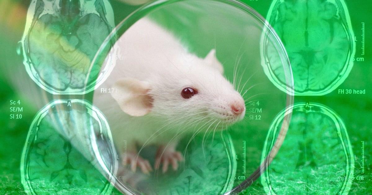
Nanoparticle Treatment Reverses Alzheimer’s in Mice
Scientists have developed a nanoparticle-based treatment that successfully reversed Alzheimer’s disease in mice.
As detailed in a new paper published in the journal Signal Transduction and Targeted Therapy, the team co-led by the Institute for Bioengineering of Catalonia, Spain (IBEC), and West China Hospital, Sichuan University, developed bioactive “supramolecular drugs” that can proactively repair the blood-brain barrier.
The barrier plays an important role in the health of the brain, defending it from harmful substances and other pathogens. Alzheimer’s has been linked to a weakening of the barrier’s integrity, allowing for impairing toxins to make it through.
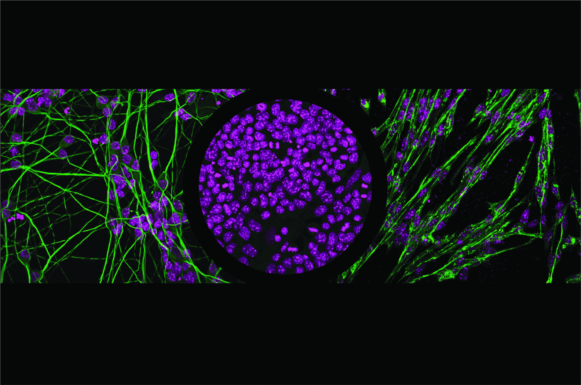
Decoding how cells choose to become muscles or neurons
Every cell in the body has the same DNA, but different cell types—such as muscle or brain cells—use different parts of it. Transcription factors help cells activate specific genes by reading certain DNA sequences, but since these sequences are common across the genome, scientists have long wondered how the factors know exactly where to bind.
Researchers in the Schübeler lab set out to address this question by looking at two closely related transcription factors—NGN2 and MyoD1—that steer cells toward becoming neurons and muscle cells, respectively. Using stem cells, they switched these transcription factors on one at a time and watched where they attached to the DNA and how they influenced gene expression. Their research is published in the journal Molecular Cell.
They found that the binding of transcription factors to the DNA molecule depends not only on the DNA sequence but also on how open the DNA is and which partner proteins are present. Sometimes, transcription factors act as “pioneer factors” and are able to open tightly packed DNA at specific sites to turn on genes. Small DNA changes—sometimes just one letter—and the proteins these factors partner with can affect whether genes are activated.
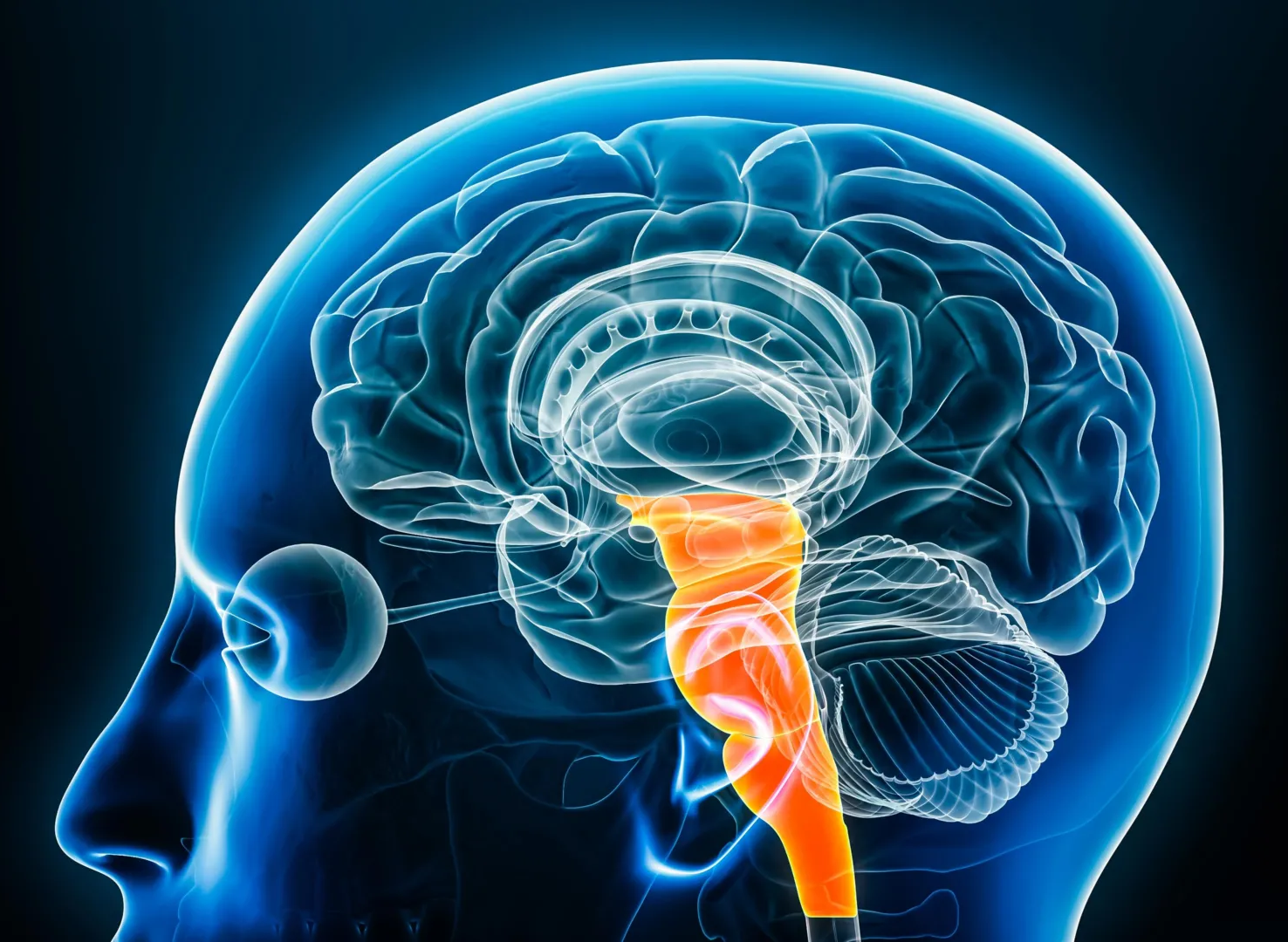
Breakthrough brain discovery reveals a natural way to relieve pain
Using powerful 7-Tesla brain imaging, researchers mapped how the brainstem manages pain differently across the body. They discovered that distinct regions activate for facial versus limb pain, showing the brain’s built-in precision pain control system. The findings could lead to targeted, non-opioid treatments that use cannabinoid mechanisms instead of opioids, offering safer pain relief options.

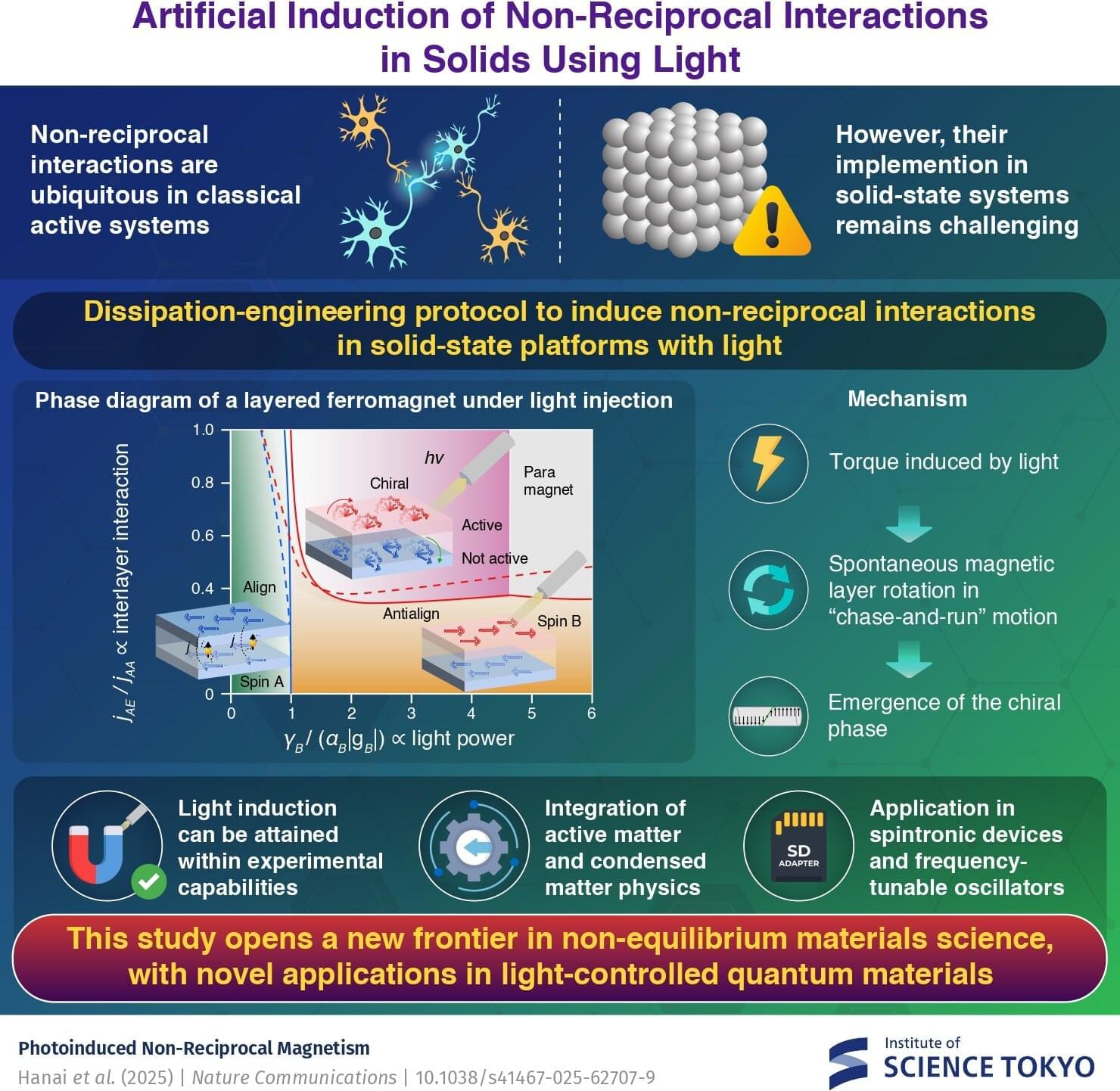
Photoinduced non-reciprocal magnetism effectively violates Newton’s third law
A theoretical framework predicts the emergence of non-reciprocal interactions that effectively violate Newton’s third law in solids using light, report researchers from Japan. They demonstrate that by irradiating light of a carefully tuned frequency onto a magnetic metal, one can induce a torque that drives two magnetic layers into a spontaneous, persistent “chase-and-run” rotation. This work opens a new frontier in non-equilibrium materials science and suggests novel applications in light-controlled quantum materials.
In equilibrium, physical systems obey the law of action and reaction as per the free energy minimization principle. However, in non-equilibrium systems such as biological or active matter—interactions that effectively violate this law—the so-called non-reciprocal interactions are common.
For instance, the brain comprises inhibitory and excitatory neurons that interact non-reciprocally; the interaction between predator and prey is asymmetric, and colloids immersed in an optically active media demonstrate non-reciprocal interactions as well. A natural question arises: Can one implement such non-reciprocal interaction in solid-state electronic systems?
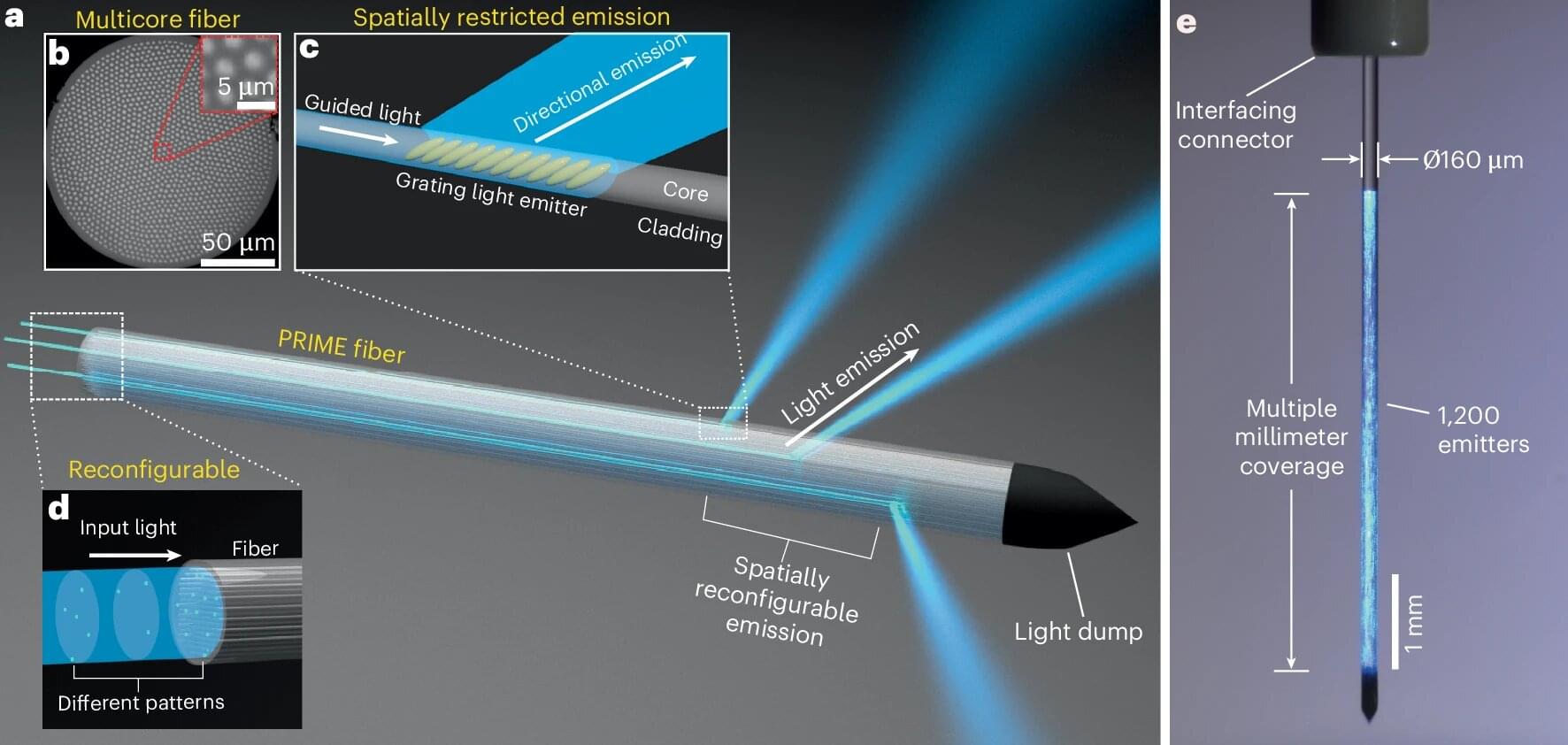
Hair-thin fiber can control thousands of brain neurons simultaneously
Fiber-optic technology revolutionized the telecommunications industry and may soon do the same for brain research.
A group of researchers from Washington University in St. Louis in both the McKelvey School of Engineering and WashU Medicine have created a new kind of fiber-optic device to manipulate neural activity deep in the brain. The device, called PRIME (Panoramically Reconfigurable IlluMinativE) fiber, delivers multi-site, reconfigurable optical stimulation through a single, hair-thin implant.
“By combining fiber-based techniques with optogenetics, we can achieve deep-brain stimulation at unprecedented scale,” said Song Hu, a professor of biomedical engineering at McKelvey Engineering, who collaborated with the laboratory of Adam Kepecs, a professor of neuroscience and of psychiatry at WashU Medicine.
The Cyborg Child: SCP-191 and the Ethics of Human Evolution
What happens when the pursuit of perfection forgets compassion?
SCP-191, known as The Cyborg Child, is one of the most haunting examples of speculative bioengineering ever documented. This essay examines the anatomy, psychology, and philosophy of a child transformed into a machine — a being caught between humanity and technology.
In this episode, we explore:
How cybernetic modification redefines the human body.
The science behind hybrid consciousness and neural integration.
The moral cost of evolution without empathy.
What SCP-191 reveals about the posthuman future.

Scientists reverse anxiety by rebalancing the brain
Researchers have discovered a specific set of neurons in the amygdala that can trigger anxiety and social deficits when overactive. By restoring the excitability balance in this brain region, they successfully reversed these symptoms in mice. The results point toward targeted neural therapies for emotional disorders. This finding could reshape how anxiety and depression are treated at the circuit level.

Rare side effects of antipsychotic medications provide new evidence for safer global prescribing
Patients with severe mental illnesses, such as schizophrenia and bipolar disorder, often require long-term use of antipsychotic medications. Some of these drugs, however, can pose potential risks, such as elevated prolactin levels and compromised immune function. Previous studies have relied mostly on small or single-center data, making it difficult to accurately assess the true incidence of rare adverse effects.
Researchers from the LKS Faculty of Medicine at the University of Hong Kong (HKUMed), through multidisciplinary collaboration and rigorous epidemiological methods, leveraged territory-wide data from the Hospital Authority to conduct two internationally impactful studies. The findings were published in the journals World Psychiatry and The Lancet Psychiatry. These discoveries provide solid evidence for drug regulation and clinical practice and establish Hong Kong as a global leader in big data research on psychiatric medication safety.