New research links multilingualism with slower aging and stronger cognitive resilience. Learn how speaking multiple languages may support long-term health.


“This Perspective concludes that an MLA between 18–21 years is a scientifically supportable and socially coherent threshold for non-medical cannabis use.”
What should be the minimum legal age for recreational cannabis? This is what a recent study published in The American Journal on Drug and Alcohol Abuse hopes to address as a team of scientists investigated the benefits and challenges of raising the legal age for using recreational marijuana to 25, with the current age range being 18 to 21, depending on the country. This study has the potential to help researchers, legislators, and the public better understand the neuroscience behind the appropriate age for cannabis use.
For the study, the researchers examined brain development for individuals aged 18–25, specifically regarding brain maturation and whether this ceases before age 25. They note it depends on a myriad of factors, including sex, geographic region, and physiology. This study comes as Germany recently published several studies regarding legalizing recreational marijuana nationwide and marijuana use rates post-legalization. In the end, the researchers for this most recent study concluded that raising the minimum legal age for recreational cannabis use to 25 is unnecessary.
The study notes, “This Perspective concludes that an MLA between 18–21 years is a scientifically supportable and socially coherent threshold for non-medical cannabis use. Policy decisions should be informed not only by neurobiological evidence but also by legal, justice, sociocultural, psychological, and historical considerations.”
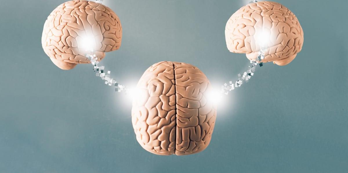
“It’s not a Jedi mind trick,” he writes in a statement. “This is what communication is. It is what humans do best, and it’s unique and amazing.”
Hasson argues that his research shows communication is really “a single act performed by two brains.” He believes that all brains naturally couple with the outside world, reacting to whatever stimuli we’re bombarded with. What makes humans different is our ability to couple without stimuli, according to Hasson. For example, if you show two monkeys a banana, their brains would likely react the same way, and the same goes for humans. However, if someone says the word banana to you, both you and the speaker would understand that you’re referring to the oblong, yellow fruit despite it not being physically present. This is something not all animals can achieve, which is why it’s so exciting for researchers like Hasson.
Studies show that brain synchronization happens in many settings. For instance, researchers found neural coupling can occur during chess matches or collaborative music-making sessions—two activities that require focus and creativity. On the other hand, a 2014 study published in PLUS One found that synchronization can occur during a much more physical activity: kissing. The experiment found heightened inter-brain connection when heterosexual couples were kissing each other’s lips rather than the backs of their hands.
The age-old advice to “trust your gut” could soon take on new meaning for people diagnosed with Parkinson’s disease, thanks to a creative feat of bioengineering by researchers in the University of Georgia’s College of Veterinary Medicine.
Anumantha Kanthasamy, professor and director of the Isakson Center for Neurological Disease Research (ICNDR) leads a multidisciplinary research team including Gregory Phillips, Piyush Padhi, and other scientists that has engineered a groundbreaking living medicine, a beneficial probiotic designed to deliver levodopa steadily from the gut to the brain of Parkinson’s patients.
In a paper published in the journal Cell Host & Microbe, Kanthasamy’s team details how they engineered and tested the probiotic bacterium Escherichia coli Nissle 1917 as a drug-delivery system that continuously produces and delivers the gold-standard Parkinson’s drug, which is converted to dopamine in the brain. The E. coli Nissle strain was chosen for its century-long record of safely treating gastrointestinal disorders in humans.

A collaboration between SISSA’s Physics and Neuroscience groups has taken a step forward in understanding how memories are stored and retrieved in the brain. The study, recently published in Neuron, shows that distinct perceptual biases—long thought to arise from separate brain systems—can, in fact, be explained by a single, biologically grounded mechanism.
The research, led by professors Sebastian Goldt and Mathew E. Diamond, and first author Francesca Schönsberg (now a junior research chair at the École Normale Supérieure), brings together theoretical physics, computational modeling, and behavioral neuroscience to bridge decades of fragmented research on perceptual memory. Yukti Chopra and Davide Giana carried out laboratory experiments to provide the empirical data that the model was tested against.

Sea urchins may just look like a ball of spikes waiting to be stepped on at the tide pool, but there’s much more to these barbed beasts than just roe and teeth.
New research reveals sea urchin nervous systems are far more complex than we knew. These creatures, it turns out, possess ‘all-body brains’ and, at least in their genetic layout, they are remarkably similar to our own.
A team of scientists led by developmental biologist Periklis Paganos from Stazione Zoologica Anton Dohrn in Italy made the discovery while investigating metamorphosis in purple sea urchins (Paracentrotus lividus), which transform from free-swimming, planktonic larvae to the mature, spine-encrusted form we’re more familiar with.

A hidden physical change in the body may be helping to drive the prolonged malaise some people experience after contracting COVID-19.
Analyzing blood samples from patients with long COVID, a team of medical researchers has identified unusual microscopic structures that may contribute to symptoms such as brain fog and fatigue. If this is the case, it offers a hopeful target for future treatment.
“This study shows a robust association between biomarkers indicative of thromboinflammatory activity and long COVID,” the team writes in a paper led by geneticist Alain Thierry of Montpellier University in France.

University of Kentucky researchers have developed a new experimental model that could point the way toward more effective Alzheimer’s disease treatments by targeting one of the brain’s most important genes for risk and resilience.
The study, published in Nature Neuroscience, focuses on apolipoprotein E (APOE), a gene long known to play a major role in Alzheimer’s disease. The team created a first-of-its-kind mouse model that allows scientists to “flip a switch,” changing the high-risk version of the gene (APOE4) to the protective form (APOE2) in adult animals.
