Scientists have uncovered a biological “blueprint” that shows how the brain’s smallest building blocks create the large networks that drive thought, emotion, and behavior.
Discover the four major turning points that reshape your brain. From childhood sharpening to late-life resilience, watch how your brain rewires itself through life. Learn the science behind cognitive peaks, aging, and brain health, and uncover the hidden timeline of your mind’s transformation. Don’t miss this deep dive into how the brain evolves over a lifetime.
#childhood #brain #science #wion.
About Channel:
WION The World is One News examines global issues with in-depth analysis. We provide much more than the news of the day. Our aim is to empower people to explore their world. With our Global headquarters in New Delhi, we bring you news on the hour, by the hour. We deliver information that is not biased. We are journalists who are neutral to the core and non-partisan when it comes to world politics. People are tired of biased reportage and we stand for a globalized united world. So for us, the World is truly One.
Please keep discussions on this channel clean and respectful and refrain from using racist or sexist slurs and personal insults.
Check out our website: http://www.wionews.com.

“Brain rot is not really rotting our brains,” says Earl Miller, a cognitive neuroscientist at Massachusetts Institute of Technology. “It’s constantly creating an environment that our brains are not equipped to deal with—that’s the real problem.
From analog hobbies to tech curfews, these Gen Zers are experimenting with science-backed ways to help their brains feel a little less foggy.
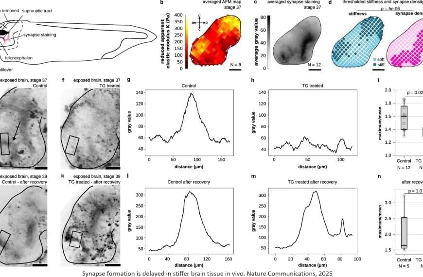
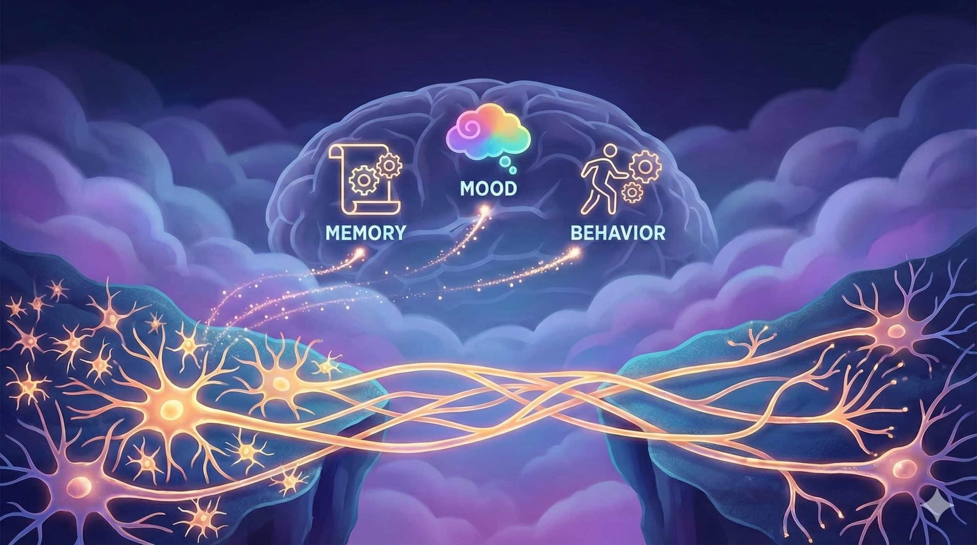
When thinking, the human brain performs a true masterpiece of information processing: around 100 billion neurons communicate with each other via approximately 100 trillion connections. An international team of researchers has discovered that the mechanical properties of the developing brain influence both synapse formation and the emergence of electrical signals. The findings could open up new approaches to understanding neurodevelopmental disorders.
In the brain, highly specific connections called synapses link nerve cells and transmit electrical signals in a targeted manner. Despite decades of research, how synapses form during brain development is still not fully understood. Now, an international research team has discovered that the mechanical properties of the brain play a significant role in this developmental process. In a study recently published in Nature Communications, the scientists showed how the ability of neurons to detect stiffness is related to molecular mechanisms that regulate neuronal development.
The developing brain is generally very soft, like cream cheese, but its stiffness varies across regions. In African clawed frog (Xenopus laevis) embryos, the researchers found that softer regions exhibit higher synapse densities, while stiffer regions show lower densities.
New research shows how human cells can be effectively ‘recharged’ by replacing their internal batteries – microscopic power stations called mitochondria – and the discovery could have wide-ranging benefits across healthcare and medical treatments.
The stacks of mitochondria in most of our cells naturally decline in numbers, slow down, and wear out with age. Once they start operating below peak capacity, they can contribute to multiple diseases everywhere from the heart to the brain.
In this latest study, researchers from Texas A&M University used special flower-shaped particles called nanoflowers to scavenge damaging oxygen molecules, triggering genes that increase the number of mitochondria in human stem cells.

Ricardo Iriart last saw his wife conscious four years ago. Every day since, he has visited Ángeles, often spending hours talking to her in hopes that she could hear him.
Over the last year, he’s gotten a new understanding of his wife’s condition, participating in cutting-edge research into “covert consciousness.” It’s an emerging field of study that probes what patients with disorders of consciousness can comprehend, even when they can’t respond.
Earlier this year, the University of Pittsburgh became the first research institution in the U.S. to use an Austrian device called the mindBeagle in a clinical trial of covert consciousness.
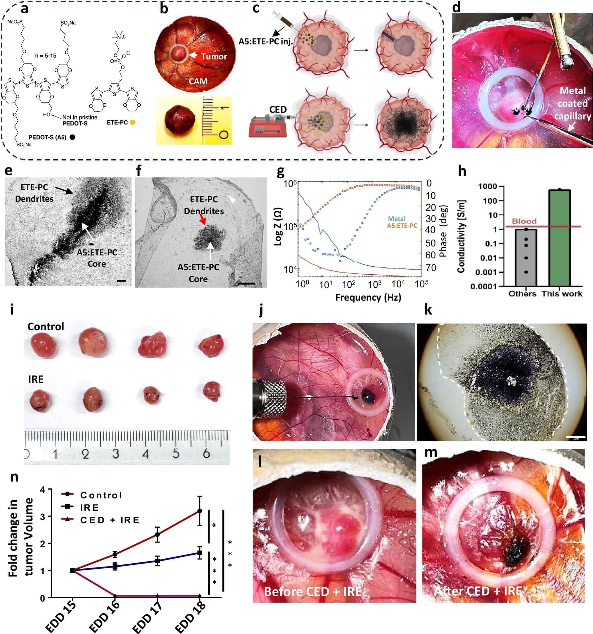
Electrotherapy using injectable nanoparticles delivered directly into the tumor could pave the way for new treatment options for glioblastoma, according to a new study from Lund University in Sweden.
Glioblastoma is the most common and most aggressive form of brain tumor among adults. Even with intensive treatment, the average survival period is 15 months. The tumor has a high genetic variation with multiple mutations, which often makes it resistant to radiation therapy, chemotherapy and many targeted drugs. The prognosis for glioblastoma has not improved over the past few decades despite extensive research.