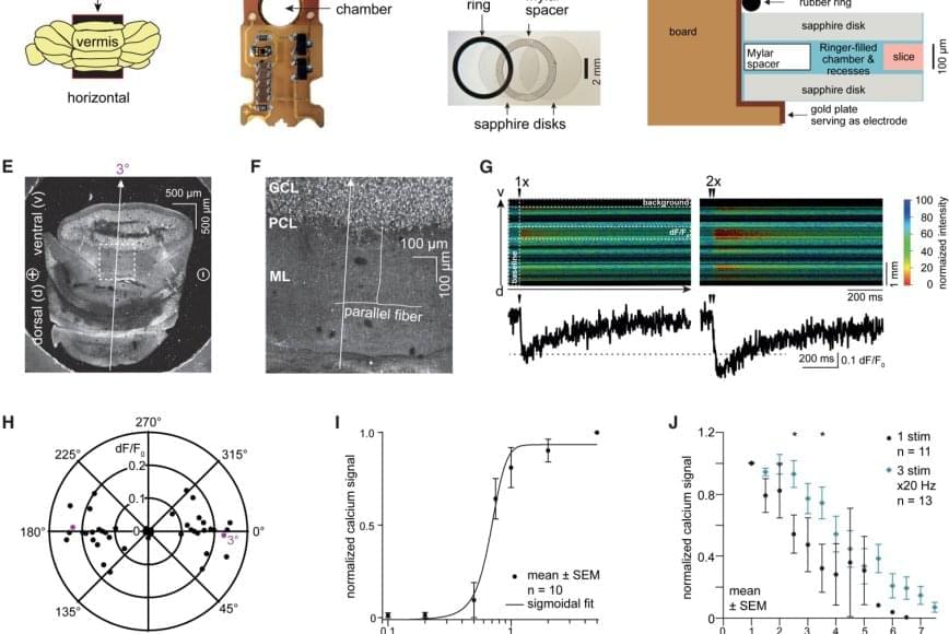
Toggle light / dark theme

Visualizing Transporter Structure Creates Platform for Antidepressant Drug Design

Researchers at Oregon Health and Sciences University’s Vollum Institute have revealed the molecular structure of the serotonin transporter (SERT), providing new insight into the mechanism of antidepressant action of two widely prescribed selective serotonin reuptake inhibitors (SSRIs) commonly used to treat depression. In their Nature paper, authors Jonathan Coleman, Evan Green, and Eric Gouaux describe their use of X-ray crystallography to capture images of human SERT structures. They collected data at the Beamline 5.0.2 in the Berkeley Center for Structural Biology and used the Phenix software suite to build models and refine the structures. The resulting structures show antidepressants citalopram and paroxetine lock SERT in an outward-open conformation, directly blocking serotonin binding.

New Ultrasound Helmet Reaches Deep Inside The Brain Without Surgery

Deep-brain structures like the basal ganglia or the thalamus wield major influence on our behavior. If something goes awry, dysregulation in the deep brain may trigger neurological conditions like Parkinson’s disease or depression.

Despite the clear importance of these structures, our knowledge about them remains limited by their location, making them difficult to study and treat.

In a new study, researchers unveil a device that might offer an alternative to invasive procedures. Featuring a novel ultrasound helmet, it not only modulates deep-brain circuits without surgery, but reportedly can do so with unrivaled precision.

Increasing the level of the protein PI31 demonstrates neuroprotective effects in mice

One fundamental feature of neurodegenerative diseases is a breakdown in communication. Even before brain cells die, the delicate machinery that keeps neurons in touch—by clearing away protein waste at the synapses—starts to fail.

When the cleanup falters, the connections between are impaired and the flow of signals responsible for reasoning, language, memory, and even basic bodily functions are progressively disrupted.

Now, a new study identifies a novel strategy for preventing unwanted proteins from clogging synapses and ultimately congealing into protein plaques.

Smells interpreted as taste!

When we eat or drink, we don’t just experience taste, but rather a ‘flavor’. This taste experience arises from a combination of taste and smell, where aromas from food reach the nose via the oral cavity, known as retronasal odor. Researchers have now shown that the brain integrates these signals earlier than previously thought – already in the insula, a brain region known as the taste cortex – before the signals reach the frontal cortex, which controls our emotions and behavior.

“We saw that the taste cortex reacts to taste-associated aromas as if they were real tastes,” explains the lead author. “The finding provides a possible explanation for why we sometimes experience taste from smell alone, for example in flavored waters. This underscores how strongly odors and tastes work together to make food pleasurable, potentially inducing craving and encouraging overeating of certain foods.”

The study involved 25 healthy adults who were first taught to recognize both a sweet taste and a savory taste through combinations of taste and smell. This was followed by two brain imaging sessions using functional magnetic resonance imaging (fMRI), in which the participants were given either a tasteless aroma or a taste without smell. The researchers trained an algorithm to recognize patterns in brain activity for sweet and savory tastes, and then tested whether the same patterns could be identified when the participants were only given aromas.

/* */