Toggle light / dark theme

Back in 1992; me and another peer who worked with me on ORNL’s ELIMS use to wish we had this technology then. And, now it looks like we’re getting closer to this capability.


An Algoma University professor has made strides in developing technology that lets ALS patients compose emails without typing.

Computer science professor George Townsend has developed the P300 Speller, a device that measures and reacts to the brain’s “surprise element” to recognize of letters of the alphabet.

The interface consists of an EEG (electroencephalogram) amplifier, an electro-cap made of spandex, and small metal electrodes that are placed over the scalp.

Read more

Big Data and Obama’s Brain Initiative — As we harness mass volumes of information and the current tech explosion around information; we will seeing an accelerated growing need/ urgency for more advance AI, QC, and new brain-mind interface intelligence to assist others when working with both super-intelligence AI and the mass volumes of information.


Engineers are experimenting with chip design to boost computer performance. In the above layout of a chip developed at Columbia, analog and digital circuits are combined in a novel architecture to solve differential equations with extreme speed and energy efficiency. Image: Simha Sethumadhavan, Mingoo Seok and Yannis Tsividis/Columbia Engineering.

In the big data era, the modern computer is showing signs of age. The sheer number of observations now streaming from land, sea, air and space has outpaced the ability of most computers to process it. As the United States races to develop an “exascale” machine up to the task, a group of engineers and scientists at Columbia have teamed up to pursue solutions of their own.

The Data Science Institute’s newest working group— Frontiers in Computing Systems —will try to address some of the bottlenecks facing scientists working with massive data sets at Columbia and beyond. From astronomy and neuroscience, to civil engineering and genomics, major obstacles stand in the way of processing, analyzing and storing all this data.

Read more

Beta rhythms, or waves of brain activity with an approximately 20 Hz frequency, accompany vital fundamental behaviors such as attention, sensation and motion and are associated with some disorders such as Parkinson’s disease. Scientists have debated how the spontaneous waves emerge, and they have not yet determined whether the waves are just a byproduct of activity, or play a causal role in brain functions. Now in a new paper led by Brown University neuroscientists, they have a specific new mechanistic explanation of beta waves to consider.

The new theory, presented in the Proceedings of the National Academy of Sciences, is the product of several lines of evidence: external brainwave readings from human subjects, sophisticated computational simulations and detailed electrical recordings from two mammalian model organisms.

“A first step to understanding beta’s causal role in behavior or pathology, and how to manipulate it for optimal function, is to understand where it comes from at the cellular and circuit level,” said corresponding author Stephanie Jones, research associate professor of neuroscience at Brown University. “Our study combined several techniques to address this question and proposed a novel mechanism for spontaneous neocortical beta. This discovery suggests several possible mechanisms through which beta may impact function.”

Read more

Many folks are not aware that one of the early detections of GBM is through a person’s weakened eyesight as well as Ophthalmologist examinations.


The retina is essentially part of the brain. Studying them led researchers one step closer to understanding how the brain processes stimuli.

There is a genetically transmitted disease that causes the eyeballs to twitch back and forth, and it’s called Nystagmus. It impacts 1 in 1,500 men. Notably, it has been recently discovered that the twitching is caused by the miscalculations done by the retinal neurons in converting visual stimuli into electrical signals.

Now, rabbits are helping us figure out how this disease operates (and could be fixed).

Read more

This amazing! I see so many uses both in medical/ healthcare as well as advancing the work in tech around brain sensory and mapping.


Sometimes it’s hard to tell the difference between science and technology ó almost all the time when it has to do with the brain. But this research from MIT that allows for vastly improved scans of the networks inside the brain is too cool to pass up, whether it’s tech, science, or somewhere in between.

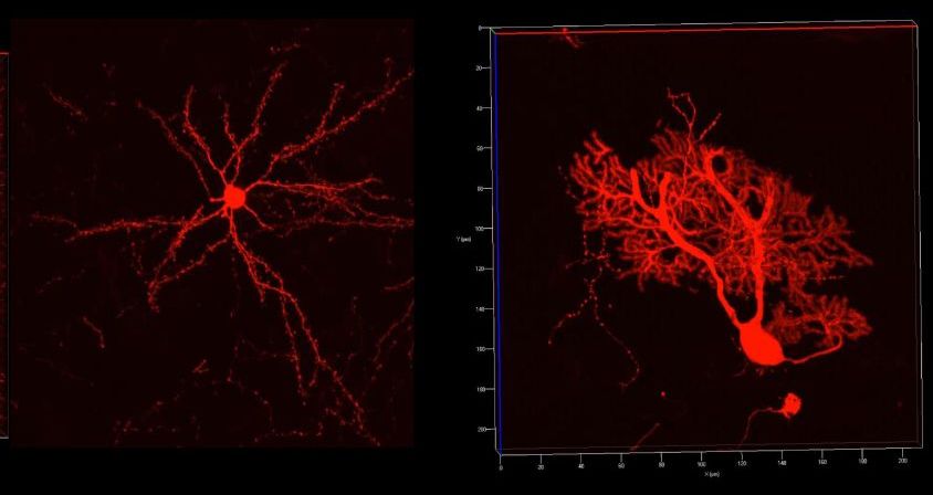
Getting up close and personal with neurons and other brain cells is a science that people have been working on for a century and more. Mainly the problem is that they’re so darn small, and packed so tightly, and connect in so many places at once, that it’s hard to tell where anything’s going. We have ways of imaging the brain at various levels, but each is highly limited in its own way.

This new technique addresses several of the main problems. It’s called magnified analysis of proteome, or (conveniently) MAP. The summary from lead researcher Kwanghun Chung makes it sound almost too good to be true.

Read more

New research sheds light on what’s going on inside our heads as we decide whether to take a risk or play it safe. Scientists at Washington University School of Medicine in St. Louis located a region of the brain involved in decisions made under conditions of uncertainty, and identified some of the cells involved in the decision-making process.

The work, published July 27 in The Journal of Neuroscience, could lead to treatments for psychological and psychiatric disorders that involve misjudging risk, such as problem gambling and anxiety disorders.

“We know from human imaging studies that certain parts of the brain are more or less active in risk-seeking people, but the neural circuits involved are largely unknown,” said Ilya Monosov, PhD, an assistant professor of neuroscience and senior author on the study. “We found a population of value-coding neurons that are specifically suppressed when animals make a risky choice.”

Read more

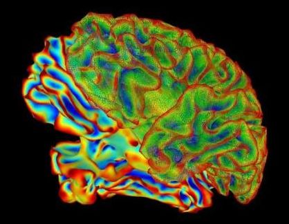
Studies are showing that anatomical patterning found in the brain’s cortex may be controlled by genetic factors.


The highly consistent anatomical patterning found in the brain’s cortex is controlled by genetic factors, reports a new study by an international research consortium led by Chi-Hua Chen of the University of California, San Diego, and Nicholas Schork of the J. Craig Venter Institute, published on July 26 in PLOS Genetics.

The human brain’s wrinkled cerebral cortex, which is responsible for consciousness, memory, language and thought, has a highly similar organizational pattern in all individuals. The similarity suggests that genetic factors may create this pattern, but currently the extent of the role of these factors is unknown. To determine whether a consistent and biologically meaningful pattern in the cortex could be identified, the scientists assessed brain images and genetic information from 2,364 unrelated individuals, brain images from 466 twin pairs, and transcriptome data from six postmortem brains.

They identified very consistent patterns, with close genetic relationships between different regions within the same brain lobe. The frontal lobe, which has the most complexity and has experienced the greatest expansion throughout the brain’s evolution, is the most genetically distinct from the other lobes. Their results also suggest potential functional relationships among different cortical brain regions.

Read more