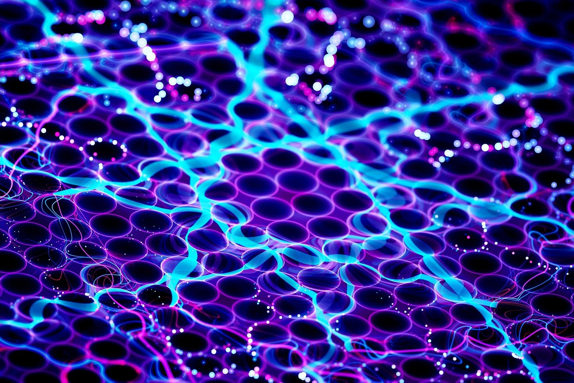
Researchers from ICMAB are revolutionizing how we manipulate light at the nanoscale using chiral plasmonic structures—nanomaterials designed to interact with polarized light in extraordinary ways.
ICMAB researchers from the NANOPTO group at ICMAB have recently published two studies demonstrating how cost-effective fabrication techniques can produce highly efficient chiral nanostructures with potential applications in sensors, imaging, and even quantum technologies.
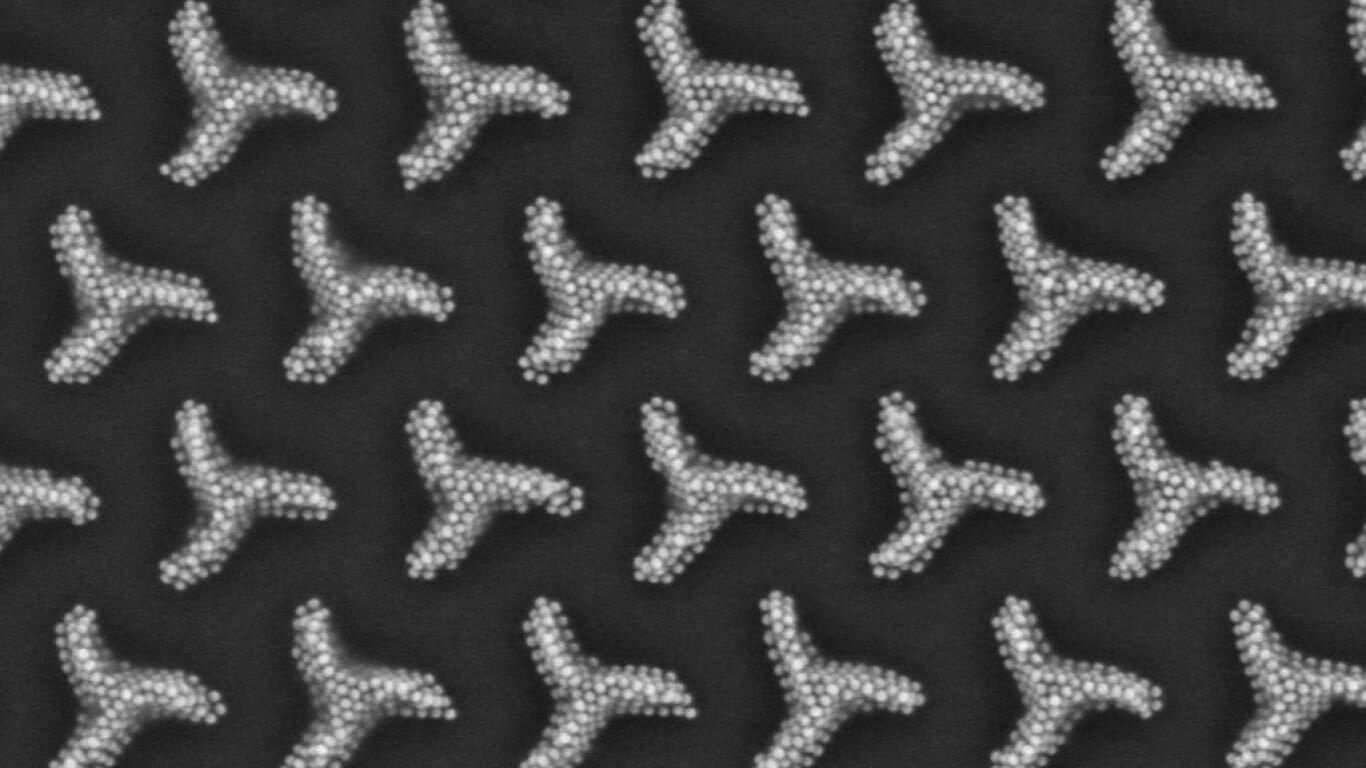
The first study, published in Nature Communications, showcases self-assembled chiral plasmonic architectures (triskelion patterns) made from gold and silver nanoparticles. These structures demonstrate exceptional optical responses, selectively interacting with circularly polarized light, opening up exciting possibilities for advanced optoelectronic devices.
MXene, a nanomaterial used in battery technology and as a high-performance lubricant, was previously difficult and hazardous to produce. However, researchers at TU Wien have now developed new, safer methods for its production. One of the most groundbreaking trends in materials science is the stud
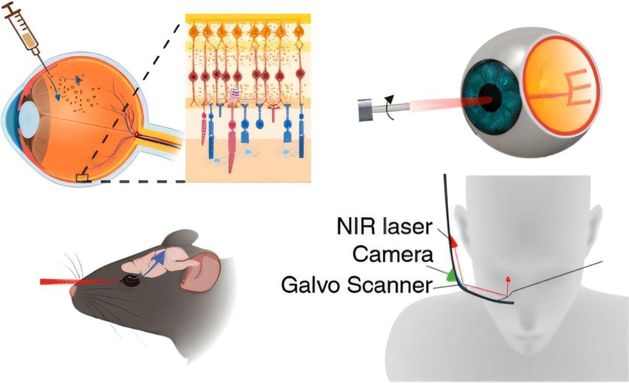
A new study by Brown University researchers suggests that gold nanoparticles—microscopic bits of gold thousands of times thinner than a human hair—might one day be used to help restore vision in people with macular degeneration and other retinal disorders.
In a study published in the journal ACS Nano, the research team showed that nanoparticles injected into the retina can successfully stimulate the visual system and restore vision in mice with retinal disorders. The findings suggest that a new type of visual prosthesis system in which nanoparticles, used in combination with a small laser device worn in a pair of glasses or goggles, might one day help people with retinal disorders to see again.
“This is a new type of retinal prosthesis that has the potential to restore vision lost to retinal degeneration without requiring any kind of complicated surgery or genetic modification,” said Jiarui Nie, a postdoctoral researcher at the National Institutes of Health who led the research while completing her Ph.D. at Brown. “We believe this technique could potentially transform treatment paradigms for retinal degenerative conditions.”
Our Vision Weekends are the annual festivals of Foresight Institute. Held in two countries, over two weekends, you are invited to burst your tech silos and plan for flourishing long-term futures. This playlist captures the magic of our Puerto Rico edition, held February 21–23, 2025, in the heart of Old San Juan. Come for the ideas: join the conference, unconference, mentorship hours, curated 1-1s, tech demos, biohacking sessions, prize awards, and much more. Stay for fun with new friends: join the satellite gatherings, solarpunk future salsa night, beach picnic, and surprise island adventures. This year’s main conference track is dedicated to “Paths to Progress”; meaning you will hear 20+ invited presentations from Foresight’s core community highlighting paths to progress in the following areas: Existential Hope Futures, Longevity, Rejuvenation, Cryonics, Neurotech, BCIs & WBEs, Cryptography, Security & AI, Fusion, Energy, Space, and Funding, Innovation, Progress. ══════════════════════════════════════
*About The Foresight Institute*
The Foresight Institute is a research organization and non-profit that supports the beneficial development of high-impact technologies. Since our founding in 1986 on a vision of guiding powerful technologies, we have continued to evolve into a many-armed organization that focuses on several fields of science and technology that are too ambitious for legacy institutions to support. From molecular nanotechnology, to brain-computer interfaces, space exploration, cryptocommerce, and AI, Foresight gathers leading minds to advance research and accelerate progress toward flourishing futures.
*We are entirely funded by your donations. If you enjoy what we do please consider donating through our donation page:* https://foresight.org/donate/
*Visit* https://foresight.org, *subscribe to our channel for more videos or join us here:*
Purdue University researchers have developed a new type of two-dimensional (2D) nanomaterial called a tungsten carbide MXene. This small but mighty material could be used to produce hydrogen fuel for electric vehicles, possibly becoming the key to a more reliable future.
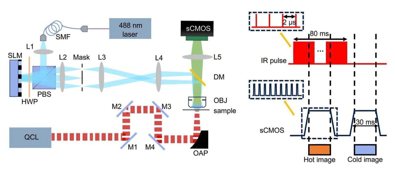
Today’s super-resolution microscopes have made it possible to observe the nanoscale world with unprecedented detail. However, they require fluorescent tags, which reveal structural details but provide little chemical information about the samples being studied.
A chemical injected before MRI scans to help create sharper images may cause some patients to experience a potentially deadly complication in rare cases, a new study suggests.
Researchers from the University of New Mexico found that gadolinium – a toxic rare earth metal used in MRI scans – could mix with oxalic acid found in many foods to precipitate tiny nanoparticles of the metal in human tissues.
The research, published in the journal Magnetic Resonance Imaging, assessed the formation of these nanoparticles associated with potentially deadly health problems in the kidneys and other organs.
Since most cells naturally repel DNA, delivering these nanodevices into cells requires specialized techniques, such as transfection methods and transformation protocols. Once inside, cellular factors such as salt concentration, molecular crowding, and heterogeneous environments influence strand displacement reactions. To overcome the limitations of direct delivery, researchers are also developing transcribable RNA nanodevices encoded into plasmids or chromosomes, allowing cells to express these circuits.
Toward Smart DNA Machines and Biocomputers
DNA strand displacement has been applied to the innovation of computational models. By integrating computational principles with DNA strand displacement, the structured algorithms of traditional computing can be combined with random biochemical processes and chemical reactions in biological systems, enabling biocompatible models of computation. In the future, DNA strand displacement may enable autonomously acting DNA nanomachines to precisely manipulate biological processes, leading to quantum leaps in healthcare and life science research.