Toggle light / dark theme

Researchers have launched a new database dedicated to mapping and understanding the complexity of cellular senescence in a bid to help us fully understand this age-related phenomenon.

Introducing the CellAge database

The Human Ageing Genomic Resources ( HAGR ) is a series of databases and tools that have been developed to aid researchers on aging and help them study the genetic elements of human aging. The databases utilize modern techniques, such as functional genomics, network analyses, systems biology, and evolutionary analyses, to build what is one of the most valuable resources available today.

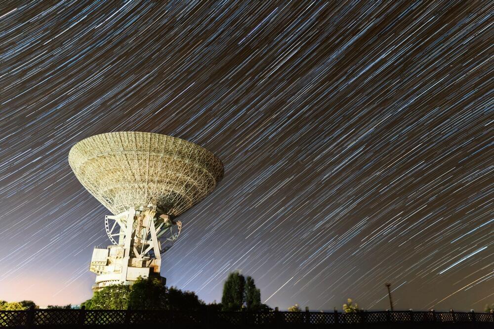
One of the biggest mysteries out there in the Universe is inching closer to answers. An astonishing eight new repeating radio signals known as fast radio bursts (FRBs) have been detected flaring from deep space.

At the start of 2019, just one of these mysterious signals, FRB 121102, was known to flash repeatedly. In January, scientists reported a second repeating one (FRB 180814).

This new paper — available on preprint server arXiv, and accepted into The Astrophysical Journal Letters — describes eight new repeating signals detected by the Canadian Hydrogen Intensity Mapping Experiment (CHIME) radio telescope.

July 20, 2019 marked the 50th anniversary of the Apollo 11 moon landing. Navy Veteran Neil Armstrong, and Air Force Veterans Buzz Aldrin and Michael Collins manned the mission.

The National Air and Space Museum displayed full-motion projection-mapping artwork on the Washington Monument. The 17 minute long show, “Apollo 50: Go for the Moon”, included a true-to-scale 363 foot Saturn V lift off, various stages of the rocket separation, the lunar landing, the first step on the moon, re-entry, and splash down back to earth.

To read more about the Apollo 11 crew, visit https://www.blogs.va.gov/VAntage/63407/veteranoftheday-apollo-11-crew/

Scientists seeking to bring to Earth the fusion that powers the sun and stars must control the hot, charged plasma—the state of matter composed of free-floating electrons and atomic nuclei, or ions—that fuels fusion reactions. For scientists who confine the plasma in magnetic fields, a key task calls for mapping the shape of the fields, a process known as measuring the equilibrium, or stability, of the plasma. At the U.S. Department of Energy’s (DOE) Princeton Plasma Physics Laboratory (PPPL), researchers have proposed a new measurement technique to avoid problems expected when mapping the fields on large and powerful future tokamaks, or magnetic fusion devices, that house the reactions.

Neutron bombardments

Such tokamaks, including ITER, the large international experiment under construction in France, will produce neutron bombardments that could damage the interior diagnostics now used to map the fields in current facilities. PPPL is therefore proposing use of an alternative diagnostic system that could operate in high-neutron environments.

In a way, the connectome is also a foundation for understanding far more complex nervous systems like our own.

“If a worm can do so much with so few neurons, and we have orders of magnitude more neurons,” Paul Sternberg, a biology professor at the California Institute of Technology in Pasadena, told Scientific American, “then we’re amazing.”

The datasets that were generated from and analysed in the current study are available at wormwiring.org

Is it possible to understand the brain? Science is still far from answering this question. However, since researchers have started training artificial intelligence on neurobiological analyses, it seems at least possible to reconstruct the cellular structure of a brain. New artificial neural networks developed by the Max Planck Institute of Neurobiology and Google AI can now even recognize and classify nerve cells independently based on their appearance.

The human brain consists of about 86 billion and about as many . In addition, there are about 100 trillion connections between the nerve cells alone. While mapping all the connections of a human brain remains out of reach, scientists have started to address the problem on a smaller scale. Through the development of serial block-face scanning , all cells and connections of a particular brain area can now be automatically surveyed and displayed in a three-dimensional image.

“It can take several months to survey a 0.3 mm piece of brain under an electron microscope,” says Philipp Schubert, doctoral student in Winfried Denk’s Department at the Max Planck Institute of Neurobiology. “Depending on the size of the brain, this seems like a lot of time for a tiny piece. But even this contains thousands of cells.” Such a data set would also require almost 100 terabytes of storage space. However, it is not the collection and storage but rather the that is the difficult part.