By genetically engineering thale cress, scientists have made it grow like a succulent, more than doubling the plant’s water-use efficiency.

There is no really useful treatments for Pancreatic Cancer, also it’s really deadly. So this sounds like awesome science news! “Cancer cells in the pancreas seem to thrive off this hyperactive cholesterol synthesis. The team thinks this is probably because they are taking advantage of other molecules generated by the same pathway. They’re able to keep the pathway running and maintain their supply thanks to an enzyme called sterol O-acyltransferase 1 (SOAT1), which converts free cholesterol to its stored form and which pancreatic cancer cells have in abundance.” “When the researchers eliminated the SOAT1 enzyme through genetic manipulation, preventing cells from converting and storing their cholesterol, cancer cells stopped proliferating. In animal experiments, eliminating the enzyme stalled tumor growth.”
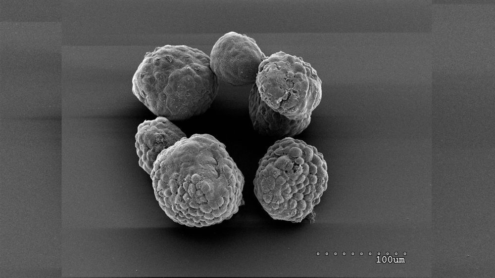
Scientists at Cold Spring Harbor Laboratory (CSHL) have found that they can stop the growth of pancreatic cancer cells by interfering with the way the cells store cholesterol. Their findings in mice and lab-grown pancreas models point toward a new strategy for treating the deadly disease.
The study, reported in the Journal of Experimental Medicine, was led by CSHL Professor David Tuveson’s team wanted to know why pancreatic cancer cells, like many cancer cells, manufacture abundant amounts of cholesterol. Cholesterol is an essential component of cell membranes, but the research team determined that pancreatic cancer cells make far more of it than they need to support their own growth. “This is unusual, because the cholesterol pathway is one of the most regulated pathways in metabolism,” says Tobiloba Oni, a graduate student in Tuveson’s lab.
Most cells make only as much cholesterol as they need, quickly shutting down the synthesis pathway once they have enough, Oni explains. But he and his colleagues, including Giulia Biffi, a former postdoctoral fellow in Tuveson’s lab, found that cancer cells convert most of the cholesterol they make into a form that can be stored within the cell. Free cholesterol never accumulates, and the synthesis pathway keeps churning out more.
Infertility is one of the most striking effects of aging. The impact of aging on females’ fertility is more severe and much better understood, but it also affects males. Male reproductive aging is less researched, but of those studies that do address it, most focus on sperm. However, ejaculate contains more than just sperm. Proteins in the seminal fluid are important for fertility, and in many animals, they have a dramatic effect on female physiology and behavior. Little is currently known about the impact of male aging on these proteins, and whether any changes contribute to poorer ejaculates in older males.
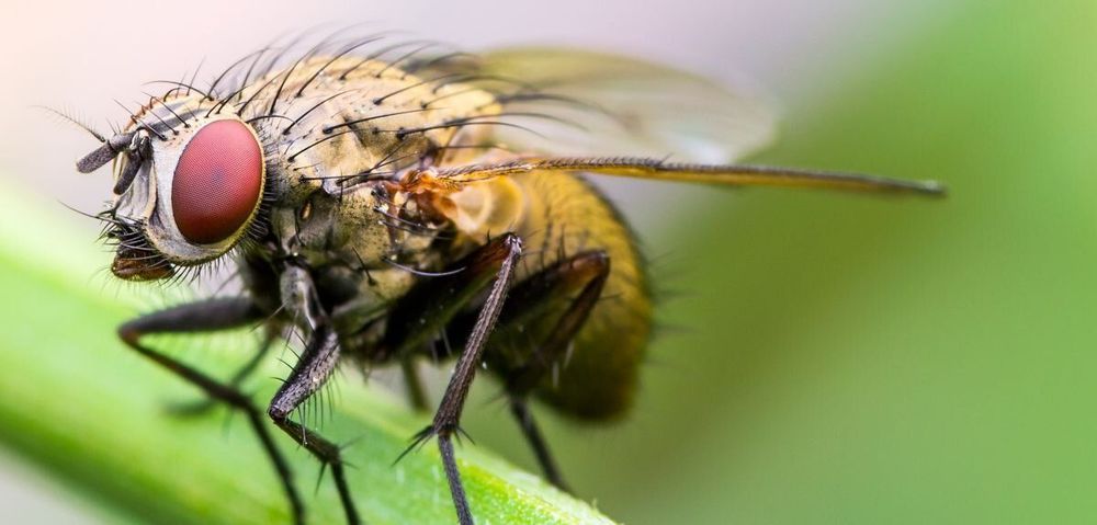
To resolve these questions, researchers at the University of Oxford’s Department of Zoology conducted experiments in a model organism, the fruit fly, Drosophila melanogaster. This species typically lives for less than five weeks, which means that researchers can very rapidly measure the impact of age on male fertility, and their sperm and seminal fluid proteins. This species is also highly amenable to genetic studies, which allowed the researchers to genetically manipulate male lifespan, to see how this impacted the decline in fertility with age.
Published this week in PNAS are their results which show that both sperm and seminal fluid protein quality and quantity decline with male age, making distinct contributions to declining reproductive performance in older males. However, the relative impacts on sperm and seminal fluid often differ, leading to mismatches between ejaculate components. Despite these differences, experimental extension of male lifespan improved overall ejaculate performance in later life, suggesting that such interventions can delay both male reproductive aging and death.

Could a mathematical model help predict future mutations of the coronavirus and guide scientists’ research as they rush to develop an effective vaccine? This is a possibility being considered by researchers at the USC Viterbi School of Engineering—Ph. D. students Ruochen Yang and Xiongye Xiao and Paul Bogdan, an associate professor of electrical and computer engineering.
Over the past year, Yang and Bogdan have worked to develop a model that could be used to investigate the relationship between a network and its parts to find patterns and make predictions. Now, Xiao is applying that successful model to the current pandemic. He is examining the RNA sequence of SARS-CoV-2, also known as coronavirus, to determine whether accurate predictions can be made about how its genetic code might change in the future based on past mutations. This research is still in progress and no conclusions have been reached yet.
Published in Nature Scientific Reports, a sister journal of Nature, Yang and Bogdan’s work is detailed in their paper, “Controlling the Multifractal Generating Measures of Complex Networks.”

Researchers from Northwest University’s medical school in Chicago believe a mutation in the coronavirus has made it considerably more contagious.
Infection disease special Egon Ozer of the Feinberg School of Medicine has said that upon examining the genetic structure of coronavirus samples, it was evident there was a change in one of the amino acids that allowed a spike in protein on the surface of the virus.
In layman’s terms, this change has allowed the virus to penetrate nearby cells easier, and as a result the virus can replicate faster and be passed on easier.

Scientists at Johns Hopkins Medicine have found types of cells in the brain that are most susceptible to inherited genetic variants linked to schizophrenia. As a result, their work reveals a shortlist of the variants that most likely impact disease risk.
Details of the scientists’ analysis, published April 17, 2020, in Genome Research, compared human genetic studies with data on how DNA is folded in mouse cells, including a diversity of brain cells.
“Every common disease has a major genetic component at its root,” says Andrew McCallion, Ph.D., professor of genetic medicine at the Johns Hopkins University School of Medicine. “Studying genomes across human populations helps us find the genetic landmarks that are linked to disease, but these often don’t give us the biological insight that pinpoints the cells in which that variation acts to impact disease risk.”

But for every insight into COVID-19, more questions emerge and others linger. That is how science works. To mark six months since the world first learnt about the disease responsible for the pandemic, Nature runs through some of the key questions that researchers still don’t have answers to.
From immunity to the role of genetics, Nature looks at five pressing questions about COVID-19 that researchers are tackling. Six months into the outbreak, Nature looks at the pressing questions that researchers are tackling.
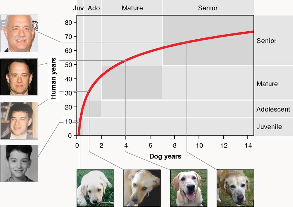
If there’s one myth that has persisted through the years without much evidence, it’s this: multiply your dog’s age by seven to calculate how old they are in “human years.” In other words, the old adage says, a four-year-old dog is similar in physiological age to a 28-year-old person.
But a new study by researchers at University of California San Diego School of Medicine throws that out the window. Instead, they created a formula that more accurately compares the ages of humans and dogs. The formula is based on the changing patterns of methyl groups in dog and human genomes — how many of these chemical tags and where they’re located — as they age. Since the two species don’t age at the same rate over their lifespans, it turns out it’s not a perfectly linear comparison, as the 1:7 years rule-of-thumb would suggest.
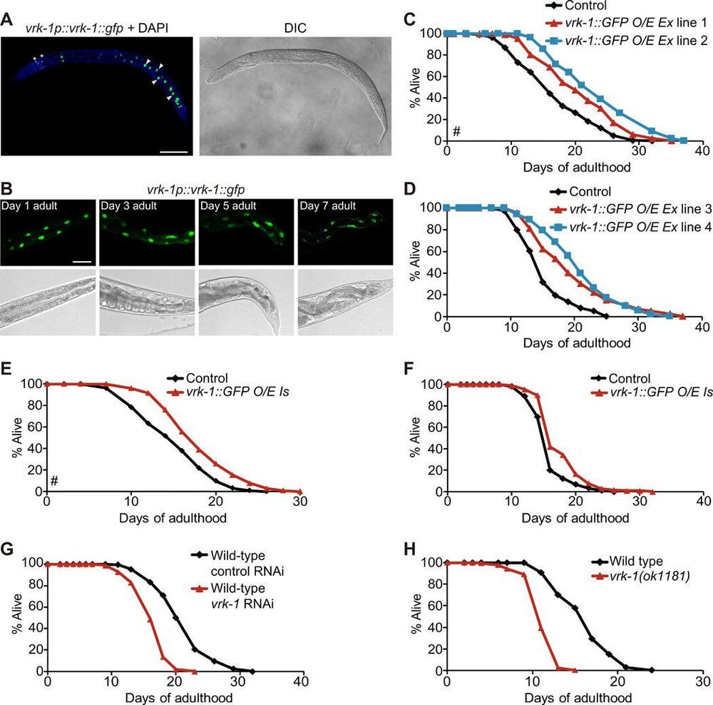
A team of researchers affiliated with several institutions in South Korea has found that stimulating production of a certain enzyme in roundworms can increase their lifespan. In their paper published in the journal Science Advances, the group describes their study of the protein VRK-1 and what they learned about its impact on the longevity of roundworms.
Prior research has shown that one way to increase longevity in some species is to use techniques that slow down mitochondrial respiration. In this new effort, the researchers were looking to better understand why slowing energy use in mitochondria has an impact on aging. As part of their effort, they looked at an energy sensor in mitochondria called adenosine 5’-monophosphate-activated protein kinase (AMPK), known to play a role in controlling how much energy is used in cells in roundworms. Prior research had suggested its level of activity is controlled by the protein VRK-1. To learn more about its impact on aging, the researchers genetically engineered two lines of roundworms to force them to produce more VRK-1 and two lines of roundworms to force them to produce less VRK-1. They then monitored the roundworms to see how long they lived.
The researchers found those roundworms expressing more than the normal amount of VRK-1 tended to live longer than average, while those expressing less than average amounts of VRK-1 had shorter lifespans. More specifically, control worms representing the normal lifespan of a roundworm lived on average 16.9 days. In their experiments, one of the lines expressing more VRK-1 lived on average 20.8 days, while the other lived on average 23.7 days. And one of the lines producing less VRK-1 lived on average just 12.7 days and the other just 15.9 days. The researchers suggest this finding indicates that VRK-1 has a direct impact on roundworm longevity.

A team of quantitative biology researchers from Northwestern University have uncovered new insights into the impact of stochasticity in gene expression, offering new evolutionary clues into organismal design principles in the face of physical constraints.
In cells, genes are expressed through transcription, a process where genetic information encoded in DNA is copied into messenger RNA (mRNA). The mRNA is then translated to make protein molecules, the workhorses of cells. This entire process is subject to bursts of natural stochasticity—or randomness—which can impact the outcome of biological processes that proteins carry out.
The researchers’ new experimental and theoretical analyses studied a collection of genes in Drosophila, a family of fruit flies, and found that gene expression is regulated by the frequency of these transcriptional bursts.